Humeral artery aneurysms (HAA) are an uncommon condition, usually secondary to trauma, infection or connective tissue disease.1,2 They have also been described in the context of arteriovenous fistulas (AVF) for haemodialysis; the majority are anastomotic or venous pseudoaneurysms, with few true degenerative arterial aneurysms. We present two cases of true HAA:
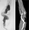
Case one is a 44-year-old male with rapidly progressive glomerulonephritis who started haemodialysis at age 20 (1978) through a left radiocephalic AVF. During the course of his illness he received two kidney transplants. In 1996 a new radiocephalic AVF was performed on the contralateral arm due to thrombosis of the previous fistula. In 2003, an asymptomatic true HAA of 6cm diameter was detected and confirmed by arteriography. Aneurysmal resection was performed with interposition of the inverted internal saphenous vein extracted from the left lower extremity. Two years later, a 4.5cm aneurysmal dilatation of the venous graft was detected (Fig. 1A) and it was repaired by PTFE prosthesis interposition. During the follow-up, no other complications were detected, with exitus in 2009 from metastatic clear-cell renal cell carcinoma.
Case two is a 55-year-old male, former smoker and with uncontrolled arterial hypertension, who started haemodialysis at age 40 (2000) through a left radiocephalic AVF due to malignant nephroangiosclerosis, receiving a kidney transplant in 2001. He had a past medical history of popliteal aneurysm in the right lower limb, operated with a femoropopliteal bypass with inverted saphenous vein, which required supracondylar amputation due to acute thrombosis. The AVF was closed in 2007 due to vein aneurysmal degeneration. In 2015, an asymptomatic true HAA of 4.5cm in diameter was documented by Doppler ultrasound and confirmed by computerised axial angiotomography (CT angiography) (Fig. 1B). Aneurysmal resection and inverted homolateral basal vein interposition were performed. At the 6-month follow-up, the bypass remained permeable and free of complications.
HAAs have been described in the context of AVF for haemodialysis, although most are anastomotic or venous pseudoaneurysms. True degenerative aneurysms are very rare, with an estimated incidence of 0.17%.1 True HAAs are most often associated with radiocephalic AVFs (60%), followed by brachiocephalic AVFs (36%), and generally appear 7–19 years after it placement.3 Unlike aortic, femoral, and popliteal aneurysms, HAAs do not appear to be associated with synchronous aneurysms in other locations.2 All of this suggests a etiopathogenic origin different from other true degenerative aneurysms.
Several mechanisms have been described that cause a significant increase in arterial diameter after the creation of an AFV; an increase in intra-arterial blood flow generates fissures in the elastic fibres in the internal lamina creating a predisposition to aneurysmal degeneration; in addition, an increase in the wall tension increases the release of endothelial factors such as nitric oxide.1,4,5 These mechanisms are neither prevented nor avoided by closure or thrombosis of the AVF.3 Kidney transplantation has been associated with aneurysmal arterial progression proximal to the AVF, while treatment with steroids and immunosuppressants has also been associated with an increased incidence of HAA.4,6,7 The two cases presented here were kidney transplant patients who received immunosuppressive therapy for more than 10 years and developed HAA 15 and 25 years after creation of the AVF, respectively.
Their most frequent clinical manifestation is in the form of an asymptomatic pulsatile mass, although they can cause pain and paresthesias due to local compression. Distal embolisation has been observed in 28–30% of cases, while the other ischaemic presentations are unusual and rupture is extremely rare.3,8,9 The initial diagnostic test is colour Doppler ultrasonography, although CT angiography is the most commonly used technique for surgical planning.10
Due to their low frequency, therapeutic indications are usually based on those accepted for popliteal aneurysms. Surgery is generally indicated for asymptomatic HAAs measuring 3cm or more. For those measuring 2–3cm, surgery is recommended if compressive symptoms or distal embolisation are present.8 First-line treatment is usually aneurysmal resection, maintaining arterial continuity by direct suture if possible. If revascularisation is required, the use of autologous grafts is preferred; if these are unavailable, prosthetic grafts or allografts would be used.3,8–10 In a systematic review published in 2015, with 12 selectable articles and 23 cases described in total, the mean permeability was 12 months (1–38 months) after autografting and 6 months (1–48 months) after PTFE interposition.10
Although systematic monitoring by ultrasound in patients with an AVF is not recommended, probably because it is not cost-effective; however it would be reasonable to perform a physical examination of the AVF at follow-up visits or at haemodialysis sessions.9 If aneurysmal degeneration is suspected it would be advisable to request a ultrasound to make early diagnosis and avoid possible thromboembolic and/or compressive complications.
Please cite this article as: Fernández Prendes C, Zanabili Al-Sibbai AA, González Gay M, Carreño Morrondo JA, Alonso Pérez M. Aneurisma humeral verdadero en relación con acceso vascular en paciente trasplantado renal: a propósito de 2 casos clínicos. Nefrologia. 2017;37:96–98.


