Introducción: la estimación del tamaño renal por ultrasonografía es un parámetro importante en la evaluación clínica y en el manejo de pacientes adultos con enfermedad renal y adultos sanos donadores. El cambio en el tamaño renal puede ser una evidencia muy sugerente de enfermedad, por lo que su interpretación requiere de parámetros específicos para la población a estudiar. En el caso de América Latina, no se han descrito parámetros normales. Objetivo: describir parámetros normales de Longitud Renal (LR) por ultrasonografía en una población mexicana adulta. Métodos: medición ultrasonográfica de LR en 153 adultos sanos estratificados por edad. Se investigó la posible asociación de la LR con parámetros antropométricos. Resultados: se estudiaron 77 varones y 76 mujeres; la edad promedio fue de 44,12 ± 15,44 años. El promedio de peso, Índice de Masa Corporal (IMC) y talla fue de 68,87 ± 11,69 kg, 26,77 ± 3,82 kg/m2 y 160 ± 8,62 cm, respectivamente. Al dividir a la población estudiada por género, encontramos que la talla fue de 166 ± 6,15 cm para varones y 154,7 ± 5,97 cm para mujeres, (p = 0,00). La Longitud Renal Izquierda (LRI) en el grupo total fue de 105,8 ± 7,56 mm, y la Longitud Renal Derecha (LRD), de 104,3 ± 6,45 mm (p = 0,000). La LRI en varones fue de 107,16 ± 6,97 mm, y en mujeres, de 104,6 ± 7,96 mm. La media de la LRD en varones fue de 105,74 ± 5,74 mm y en mujeres, de 102,99 ± 6,85 mm, (p = 0,008). La LR disminuyó con la edad, y la tasa de disminución parece aumentar después de los 60 años. Las LR se correlacionaron de forma significativa y positiva con el peso, el IMC y la talla. Conclusiones: la LR fue significativamente mayor en varones que en mujeres para ambos riñones (p = 0,036). La LR disminuyó continuamente con la edad, especialmente después de los 60 años y de forma significativa después de los 70 años.
Introduction: Renal length estimation by ultrasound is an important parameter in clinical evaluation of kidney disease and healthy donors. Changes in renal volume may be a sign of kidney disease. Correct interpretation of renal length requires the knowledge of normal limits, these have not been described for Latin American population. Objective: To describe normal renal length (RL) by ultrasonography in a group of Mexican adults. Methods: Ultrasound measure of RL in 153 healthy Mexican adults stratified by age. Describe the association of RL to several anthropometric variables. Results: A total of 77 males and 76 females were scanner. The average age for the group was 44.12 ± 15.44 years. The mean weight, body mass index (BMI) and height were 68.87 ± 11.69 Kg, 26.77 ± 3.82 kg/m2 and 160 ± 8.62 cm respectively. Dividing the population by gender, showed a height of 166 ± 6.15 cm for males and 154.7 ± 5.97 cm for females (p =0.00). Left renal length (LRL) in the whole group was 105.8 ± 7.56 mm and right renal length (RRL) was 104.3 ± 6.45 mm (p = 0.000). The LRL for males was 107.16 ± 6.97 mm and for females was 104.6 ± 7.96 mm. The average RRL for males was 105.74 ± 5.74 mm and for females 102.99 ± 6.85 mm (p = 0.008). We noted that RL decreased with age and the rate of decline accelerates alter 60 years of age. Both lengths correlated significantly and positively with weight, BMI and height. Conclusions: The RL was significantly larger in males than in females in both kidneys (p = 0.036) in this Mexican population. Renal length declines after 60 years of age and specially after 70 years.
INTRODUCTION
Renal length estimation by ultrasound is an important parameter in clinical evaluation of adult patients kidney disease and healthy adult donors1,2 and has replaced radiography as the common standard. Ultrasound is a useful, accessible, non-invasive, inexpensive method to reliably measure renal size.3
Some renal diseases can change the morphological characteristics of the kidney seen by ultrasound. Renal size can also be a decisive factor for performing renal biopsy or avoiding immunosuppressive therapy.2 Estimating renal size by ultrasound can be done by measuring the length, total volume or cortical thickness. The most accurate measurement of renal size is the total renal volume, which is correlated with height, weight and total body area. This measurement requires expensive, highly complex studies with specific protocols, such as axial tomography and magnetic resonance. However, renal length has also been shown to be a reliable parameter2 with a high level of interand intra-observer reproducibility in comparison to volumetric renal estimation, which correlates appropriately with function and different anthropometric variables.4
Renal size depends on different factors, which include size, body mass index and gender. However, race has particular connotations, which directly determines all the previous variables. The change in renal size can be very suggestive evidence of disease, whose interpretation requires specific parameters for the population to study. It is therefore necessary to have benchmark parameters in our population group.
MATERIALS AND METHODS
Prospective observational study carried out during the period between April and June 2008. An ultrasound screening was carried out on 153 healthy volunteers who complied with the following criteria for inclusion: serum creatinin ≤ 1.5mg/dl, glycaemia ≤ 110mg/dl in patients aged over 40 years or with BMI > 30kg/mt2, arterial normotensive (systolic blood pressure < 140mmHg and diastolic blood pressure < 90mmHg), no existence of acute or chronic disease capable of causing damage to renal function and normal appearance of the kidneys by ultrasound (thickness of renal parenchyma > 1cm and corticomedullary ratio detectable by ultrasound.) Patients with the following conditions were excluded: cysts greater than 4cm, polycystic kidney disease, multiple cysts (> 4), sole kidney, hydronephrosis, poor ultrasound examination window (automatically elevated kidney, with interference in costal arches), pregnancy, extreme obesity, renal tumours and horseshoe kidney.
Height was measured without shoes or hat, using a stadiometer. The ultrasound measurement was made by a single observer, using a TITAN Sonosite high resolution device, with a 3.5MHz convex transductor. All the participants emptied their bladders prior to the examination, to avoid an increase in renal length caused by oral hydration.5 Renal length was measured as the longest longitudinal diameter, with the patient fasting and in three positions (supine, supine lateral and prone.) Three measurements were taken for each kidney, registering the longest length in absolute terms.
The results were expressed as an average + DS. The averages of the different numerical variables were compared by gender using the t-Student test for independent samples. An association test was performed between the different renal lengths and the anthropometric variables using the Pearson correlation coefficient. Finally, the total student population was divided into age groups with 10-year intervals and a comparison was LRL and RRL between the different age groups using a one-way analysis of variance, followed by Bonferroni’s multiple-comparison test (post hoc.) A value of p < 0.05 was considered statistically significant. The SPSS version 15 statistical packet for Windows was used.
RESULTS
A total of 157 subjects were included, four of whom were excluded due to the presence of solid cysts over 4cm long (two subjects), morbid obesity (one subject) and one case with a very thin renal cortex (< 1cm). Of the 153 individuals evaluated, 77 were male and 76 female, with an average age of 46.12 + 15.44 years (range between 20 and 79 years.) The average for the different anthropometric measurements in the total study population (weight, height and BMI) was: 68.87 ± 11.69kg, 160 ± 8.62cm and 26.7 ± 3.82kg/mt2, respectively. The left renal length in the total group was an average of 105.8 ± 7.56mm, and the right renal length 104.3 ± 6.45mm (p = 0.000.) With regard to the Cr S, the average value for the total group 0.86 ± 0.17, with a range of 0.5 to 1.3mg/dl; and specifically for the 60 to 70 years age group the average value was 0.94 ± 0.17, with a range of 0.63 to 1.3mg/dl. The glomerular filtration for this age group, estimated with the Cockroft-Gault formula, corresponds to 66 ± 14.8ml/min, with a range of 42 to 93ml/min.
When the study population was divided by gender, the following data was found: the average weight was 73.77 ± 11.29kg for males (range of 52 to 111kg) and 63.9 ± 9.90kg for women (range of 43.5 to 85kg) (p = 0.00.) The average height was 166 ± 6.15cm for males (range of 155 to 185cm) and 154.7 ± 5.97cm for women (range of 139 to 167cm) (p = 0.00.)
The average LRL in men was 107.16 ± 6.97mm (range of 90 or 121mm), and in women, from 104.6 ± 7.96mm (range from 88 to 122mm) (p = 0.036.) The average RRL in men was 105.74 ± 5.74mm (range of 93 or 120mm), and in women, from 102.99 ± 6.85mm (range from 89 to 120mm) (p = 0.008.)
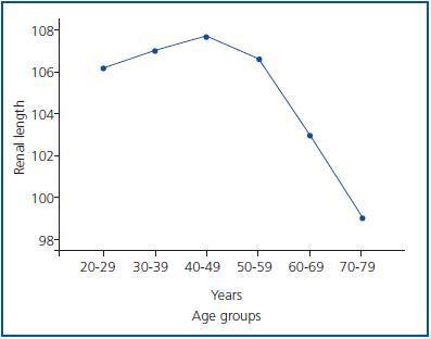
When the total study population was divided up according to age groups (group 1: 20-29 years; group 2: 30-39 years; group 3: 40-49 years; group 4: 50-59 years; group 5: 60-69 years; group 6: 70-79 years), and when their LRL are compared, the following measurements were found by group: 106 ± 6.53mm (range from 95 to 119mm), 106.9 ± 6.20mm (range from 93 to 119mm), 107.6 ± 8.3mm (range from 91 to 122mm), 106 ± 6.9mm (range from 92 to 120mm), 102.9 ± 8mm (range from 88 to 116mm) and 99 ± 7.92mm (range from 89 to 118mm) from groups 1 to 6, respectively, significantly different in form for 6 vs. groups 2 and 3 (p < 0.05) (table 1 and figure 1.)
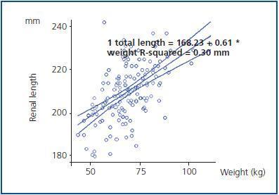
With regard to the RRL, the following measurements were observed per group: 103 ± 6.06mm (range from 92 to 120mm), 105 ± 5.57mm (range from 94 to 116mm), 105.8 ± 7mm (range from 90 to 120mm), 106 ± 6.0mm (range from 95 to 116mm), 102 ± 6.5mm (range from 92 to 114mm) y 100 ± 6.93mm (range from 89 to 110mm) for groups 1 to 6, respectively, which were not significantly different (p = NS) (table 1.) The association test performed using the Pearson correlation coefficient showed a significant positive correlation between both renal lengths and the different anthropometric measurements (weight, BMI and height), for the LRL (r=0.516, p=0.000; r = 0.408; p = 0.000; and r = 0.260, p = 0.001, respectively) and for the RRL (r = 0.501, p = 0.000; r = 0.363, p = 0.000; and r = 0.289, p = 0.000, respectively) (table 2 and figure 2.)
DISCUSSION
Renal disease can increase or decrease renal size, and may or may not be accompanied by changes to the normal organ structure. Ultrasound has been shown to be a diagnostic method for taking these measurements, which offers the advantage of being a non-invasive, innocuous method for the patient in comparison to other measurement methods such as simple radiography or intravenous urography, which have also shown their effectiveness in renal size evaluation.
Renal length and volume are important parameters in the clinical scenario. Specifically, renal length measurement is more valuable in adults due to its reproducibility and accuracy. Because of this, it is essential to know the normal limits of renal size in patients for the purposes of correctly interpreting the study.
In this study a significant difference in weight and height was observed when gender comparisons were made (p = 0.00.) At the same time, gender-related differences related to the RL were confirmed, showing that the length of both kidneys was significantly greater in men than in women. Similar data to that shown in earlier studies has been shown,8,9 in contrast to the paediatric population, where generally speaking no significant differences have been seen in renal length, probably related to the size of the sample and the strength of each study.10
It is of great relevance that the decrease in both renal lengths (LRL, RRL) from 60 years, which was significant from 70 years on, mainly for the LRL (p < 0.05), while the renal lengths in subjects aged between 20 and 59 years remained relatively homogeneous. Therefore, it would appear that renal length decreases considerably with age and that the rate of decrease accelerates with age, especially after 60 years, but above all after 70 years. Other authors have shown results, showing that the length and renal volume in subjects aged between 30 to 60 years showed slight differences and a clear decrease in both parameters after 70 years.9 In this context, the changes in renal structure and shape that take place in human beings with age, decreased renal mass and renal weight and size, are well known.11 Post mortem studies have shown that the weight of the kidney is between 250 and 270g in young adults, which falls over the years to 200g in the ninth decade of life.12 Factors such as glomerulosclerosis and tubulointerstitial fibrosis could lead to a decrease in renal size and weight, as histological data reveals a decrease in the number of cortical glomerules by 30-50% at the age of 70 years, as well as observing a loss of glomerular lobulation, increasing mesangial volume and glomerular collapse, as well as intimal thickening hyalinosis of both the arteries and the arteriola.13-15 The latter can be explained by a decrease in the proangiogenic vascular endothelial growth, as well as an increase in the expression of the antiangiogenic factor thrombospondin 1, as has been shown in elderly rats. Among other factors associated as potential modulators of renal fibrosis in elderly animals, angiotensin II has been mentioned, which is the beta transformer growth factor (TGF-β), changes in the expression de Nitrous Oxide (NO), final products of advanced glycosylation (AGE), oxidative stress and changes in lipids, may be future targets to modulate the progression of sclerosis and the decrease in renal size.12
The decrease in renal length at 60 years, and in particular from 70 years, was significant for LRL, not for RRL, which showed no significant difference between the age groups in our study, in contrast to the results published by other authors. The left kidney was significantly larger than the right, which was similar to the results of the majority of studies published. This difference could be explained by the fact that the liver is bigger than the spleen, which means that the right kidney has less space to grow. Another possible explanation is related to the shorter length of the left renal artery, which increases blood flow to the left kidney, with the corresponding increase with regard to the volume and size of the kidney. Finally, the height (shorter in our population) may limit the free longitudinal growth of both kidneys.
On the other hand, the two renal lengths correlate in a significantly positive manner with weight and BMI, and weakly with height, in contrast to other studies in adults where the strength of this association has been significantly greater with height.8,9 On the other hand, in the paediatric population a greater association with renal length has been found with the average TFG with inulin6 and with body surface area.16
We consider it pertinent to explore the association of renal lengths in the future, or their sum (total renal length), with different anthropometric measurements, as well as with renal function parameters.
CONCLUSIONS
In conclusion, LRL was significantly greater than RRL in our population, and both renal lengths were greater in men than in women. Renal size and height were shorter in this population in comparison with figures published in European populations. A fall in the RL was observed with age, especially after 60 years, but significantly after the age of 70 years. Both RL correlated positively with weight, the BMI and height.
Figure 1.
Table 1. Distribution of renal length according to age group (years)
Figure 2.
Table 2. Correlation of anthropometric measurements with renal length


