Se estudian tres pacientes con enfermedad renal crónica en terapia hemodialítica, en los cuales se habían agotado los accesos venosos clásicos en el hemitórax superior (yugular interno, subclavio, axilar) para hemodiálisis, debido principalmente a trombosis de los mismos por cateterismos anteriores. En ellos se optó, mediante la técnica de Rao et al., por puncionar la vena innominada, lográndose la implantación posterior de catéteres y su tunelización subcutánea. Los catéteres permanentes3 funcionaron adecuadamente y están permeables a la fecha después de un periodo promedio de ocho meses.
INTRODUCTION
Vascular accesses represent a serious problem in patients with chronic kidney disease (CKD) who are undergoing haemodialysis. Arteriovenous fistulae (AV) with native vessels or grafts are ideal accesses because of their long life and low rate of complications during use. In patients for whom it is not possible to create an AV fistula, it is necessary to implant central catheters in order to carry out the haemodialysis treatment. However, during their placement and use, these catheters generate various complications,1 one of which is thrombosis of the vessels in which they are inserted. This means that with the passing of time, patients run out of conventional vessels (internal jugular, subclavian, axillary vein) in the upper hemithorax, and it becomes necessary to access the superior vena cava through uncommonly used vessels in order to perform haemodialysis. In this article, we present an alternative approach to the placement of haemodialysis catheters in the brachiocephalic vein, also known as the innominate vein, in patients with a history of thrombosis in the more conventional vessels.
CASE DESCRIPTION
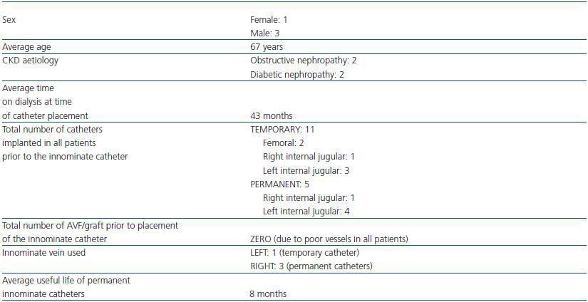
The patients selected for catheter placement in a brachiocephalic vein were those who had catheters previously inserted in their internal jugular and axillary veins and in whom ultrasound showed those sites to be thrombosed, whether due to previous catheterisation or to congenital malformation. All of the patients gave written consent to have the procedure performed; exclusion criteria were coagulation test abnormalities (PTT and PT), thrombocytopenia (a platelet count below 50,000 platelets) or refusal to accept the procedure. The characteristics of the selected patients are shown in table 1.
The inclusion criteria are based on our Kidney Unit¿s protocol for the insertion of central venous catheters in which the preferred site is the right jugular vein first, followed by the left jugular vein and finally the axillary veins. We do not generally puncture the subclavian vein because of the risk of complications as explained below.
All the patients had an initial ultrasound scan of the neck vessels and the infraclavicular region to exclude the existence of thrombosis in both internal jugular veins, the subclavian and axillary veins and this was consistently documented. For all patients, we were able to determine the location of the brachiocephalic vein by placing the transducer just above the clavicle.
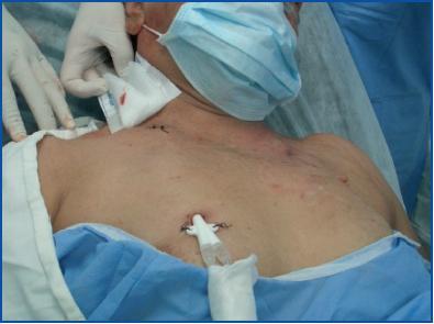
Patients were positioned supine and the supra- and infraclavicular regions were cleaned. Using aseptic technique and local anaesthetic, the needle was introduced with sustained aspiration directly above the clavicle between the sternal and clavicular heads of the sternocleidomastoid muscle, toward the mediastinum and parallel to the anterior thoracic wall, until obtaining a good flow of blood. We achievement puncture the vein easily, 2-4cm from the skin puncture site; once the vein had been reached, we proceeded to thread the guidewire, dilate the tunnel and place the permanent catheter. It is then tunnelled through and brought out the right hemithoracic wall.
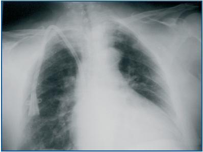
Three patients had four catheters placed in the innominate vein: three on the right side (all permanent), and one on the left side, which was temporary. No complications arose that could be attributed to the procedure.
The temporary catheter was removed 15 days after its insertion due to it not functioning properly; the three permanent catheters continue to work properly to date, an average of eight months after their implantation.
DISCUSSION
The implantation of central catheters is a routine procedure for most nephrologists. The internal jugular vein approach is the most commonly used, due to the ease of cannulation and the low rate of complications.2 The subclavian route is not recommended because of its propensity for stenosis and thrombosis, which subsequently prevent use of the ipsilateral side for AV fistulae creation.3 The axillary vein may also be used for the placement of central catheters, but reaching this vein requires medical personnel familiar with the cannulation procedure.4 The femoral route is easy to access, but its drawbacks are high rates of thrombosis and infections5 making this site unsuitable for long-term catheter placement. Other routes that are used in special situations are the transhepatic and translumbar routes.6,7
One of the complications associated with long-term use of central venous catheters is vein thrombosis and stenosis. Various alternatives have been suggested for recanalization of occluded vessels, including implanting stents8 and prostheses that replace stenosed segments,9 but these are associated with a high rate of re-occlusion a few months later. This fact obliges us to explore other routes to guarantee that haemodialysis patients have a satisfactory access, and the innominate vein is an alternative that is not often used.
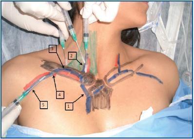
It is important that we do not confuse the approach to the innominate vein with the supraclavicular approach used to reach the subclavian vein. This is done by puncturing directly above the clavicle, but on the external side of the clavicular head of the sternocleidomastoid (figure 1) in the medial direction, requiring patency of the subclavian vein.10,11
We recommend puncturing the right innominate vein, given that in the left hemithorax the pulmonary cupola is higher and the thoracic duct crosses directly over those planes intersected by the needle, which exposes the patient to complications such as pneumothorax and chylothorax.
In 1977, Rao et al.12 published their experience with developing a new technique for placing central venous catheters using the innominate vein.
The technique of placing catheters in the innominate vein has been performed successfully in patients with thrombosis or stenosis of the internal jugular and subclavian veins, being widely used for anesthesiologists, and has become the main route for venous access in some medical centers.13
For patients with chronic kidney disease who undergo haemodialysis treatment, few studies exist in the international literature regarding the use of the innominate vein for vascular access. The first report was presented by Apsner et al.14 in 1996, with four catheters being inserted in four patients, all of whom had similar characteristics to those we describe. Lau et al.15 described a further two cases in 2001, and the largest study led by Falk A was published in 2006,16 in which 44 innominate vein catheters were placed in 33 patients, importantly without serious complications during the procedure and with long-term patency.
In conclusion, approaching the superior vena cava through the innominate vein is an alternative route, but with practice, it can come to represent an excellent option for patients with multiple thrombosed veins in their upper hemithorax.
Figure 1.
Figure 2.
Figure 3.
Table 1. Characteristics of patients selected for catheter placement in the innominate vein


