To the Editor,
Peritonitis is the primary cause of morbidity, mortality, and technique failure in peritoneal dialysis (PD).
Several studies have shown that catheter removal (CR) is necessary in as many as 16%-18% of cases.1 The most common causes of peritoneal CR due to peritonitis are: fungal peritonitis, enteric peritonitis, and cases associated with other clinical circumstances (simultaneous infection of the subcutaneous tunnel, cases refractory to treatment, and recurring infections).
Following CR, a high percentage of patients decide to stay with the same method of depuration treatment. These patients tend to have a low technique survival due to adherence and ultrafiltration failure.2
If patients decide to reinitiate PD, it is important to keep in mind:
1. There is no reliable, objective method that can indicate the existence of peritoneal damage before inserting a catheter: ultrasound, computed tomography (CT), and magnetic resonance (MRI) have all been shown to have low sensitivity and only provide imprecise detection of alterations.3
2 The catheter should be inserted under open or laparoscopic surgery in order to obtain visual information about the abdominal cavity.
3, The new catheter should be inserted a minimum of 3-4 weeks after the complete recovery from the infection.
4. In the case of early catheter dysfunction, a peritoneography can be useful in the chance of compartmentalisation of the peritoneum.
We carried out a retrospective study during the last five years on patients in our unit that required CR because of peritonitis and that later decided to reinitiate PD.
CR was required in 11 patients from our study population following cases of peritonitis in the last five years.
We performed abdominal CT scans for each prior to inserting the second catheter.
Only one patient was denied reinitiation of PD when the CT scan revealed an abdominal image indicative of a small abscess two months after the removal of the first catheter.
The second catheter was inserted in all cases under general surgery conditions; mild adherence was observed in two cases, which were remedied.
The mean age of patients in our study was 62.8 years (range: 30-77).
Mean albumin levels were 3.5mg/dl (2.8-4.2); mean D/P creatinine at 240 minutes: 0.75 (0.69-0.8); mean D/P creatinine 240 minutes before removal: 0.78 (0.63-0.9); mean total number of cases of peritonitis per patient: 2.6 (1-5), and the mean time until the appearance of the first case of peritonitis was 18 months (1-47).
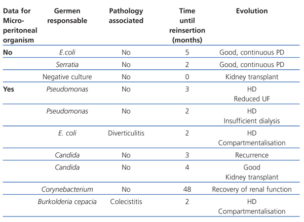
The micro-organisms responsible for the cases of peritonitis, the existence of associated pathologies, the time until reinsertion of the second catheter, and patient evolution (resolution or lack thereof of the infectious problem before the CR) are summarised in Table 1.
In our study sample, almost all patients whose catheters were removed during the infection had poor evolution of the dialysis technique, primarily due to adherences or problems with ultrafiltration, similar to the results from other studies.4
Although imaging tests prior to the second catheter insertion have low sensitivity, we believe that they are necessary, since an abdominal pathology secondary to the first case of peritonitis may be present, with no clinical symptoms, as occurred in our case of a patient with an abdominal abscess.
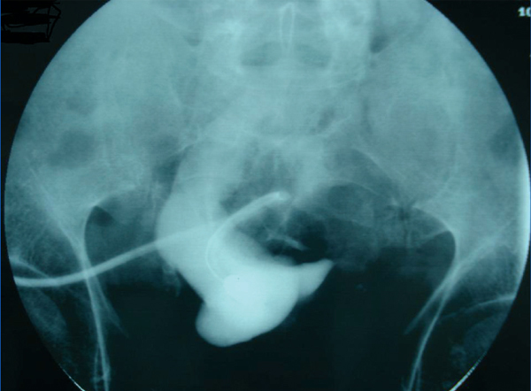
In the case of early dysfunction of the peritoneal catheter, a peritoneography is necessary for evaluating the presence of compartmentalisation (Figure 1).
In conclusion, a return to PD following CR due to peritonitis should be evaluated on an individual basis, paying special attention to those patients that had peritonitis refractory to treatment, with associated abdominal pathologies, and a high D/P creatinine level before the removal of the first catheter. The impact that the possible loss of residual diuresis would have on the evolution of the patient should also be taken into account.
Conflicts of interest
The authors have no potential conflicts of interest to declare.
Table 1. Causative micro-organisms and patient evolution following removal of the peritoneal dialysis catheter
Figure 1. Image of a pseudocavity in the peritoneography


