To the Editor,
Cystinuria is a hereditary disease caused by a defect in the renal and intestinal tubular transport affecting cystine and the dibasic amino acids (lysine, ornithine and arginine).1 It is transmitted as an autosomal recessive disorder and has a prevalence of about 1 in 7,000 live births, with a wide geographical variation and no predominance of sex. The clinical manifestations are effectively nephrolithiasis and its consequences (colic, haematuria, etc.) which usually occur in the second or third decades of life, although they can appear as early as the first year. It is the cause of 6%-10% of paediatric urolithiasis cases.2 The cystine stone formation is due to the excessive concentration of this amino acid in urine and its high insolubility, especially when the urine is acidic.
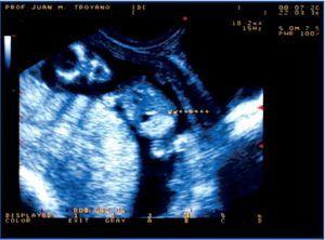
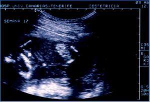
We had the opportunity of studying a child, currently three years old, who was referred by his paediatrician when he was five months old, after an episode of gross haematuria, which revealed the presence of a stone in the nappy. It was the first child of non-consanguineous parents, without any previous significant pathology, but with a history of renal colic on the paternal side of the family. Ultrasound foetal studies during pregnancy revealed a colon hyperechogenicity without other intestinal abnormalities (Figures 1 and 2), and a slightly increased nuchal luminescence, with no other findings of interest. As a result, a sweat test was performed at birth to rule out cystic fibrosis and the result was normal.
Subsequent ultrasound images revealed multiple bilateral stones, which grew to a diameter of 1.4cm. Persistently high cystine elimination was detected in the urine (maximum 656mg/g creatinine at 7 months old). The renal glomerular function is normal (serum creatinine 0.28mg/dl), although there was a defect in the ability to concentrate (689mOsm/kg) and elevated urinary excretion of microalbumin (microalbumin/creatinine ratio 33.9μg/μmol).
During its evolution, numerous small stones have been expelled (over 50 during the first year of life, measuring few mm in diameter), and the condition is otherwise asymptomatic. The weight-to-height ratio and psychomotor development during growth was normal. Pharmacological and dietary treatment with potassium citrate, captopril and D-penicillamine is currently being administered.
This is an early clinical presentation of cystinuria, reflecting the high lithogenic capacity of this condition. The particularity of the case is that the prenatal ultrasound found hyperechogenicity of the colon secondary to cystine crystal deposition. This form of presentation of cystinuria was described in 20063 and was subsequently confirmed.4 The explanation for this finding is that the cystine crystals are formed in the foetal kidney, they enter the amniotic fluid and are then swallowed. The ultrasound finding of the foetal hyperechogenic colon has been traditionally related to cystic fibrosis, which was why the studies needed to rule out the disease were performed at birth. The negative result and early clinical symptoms led to the diagnosis. Knowledge of this association may facilitate an early diagnosis of the disease, thus establishing an appropriate treatment.
Figure 1. Hyperechogenic intestine with sound density similar to foetal bone
Figure 2. A similar situation early in the second trimester


