To the Editor,
Aortocaval fistula (ACF) is an uncommon complication of abdominal aortic aneurysms (AAA) that requires urgent action. However, they are diagnosed in only half of cases, which increases postoperative mortality. Indicative of this condition are a continuous abdominal murmur and the presence of high-output heart failure (HF) or regional venous hypertension. Acute renal failure (ARF) may be the presentation of the ACF, as in the case described.
This is the case of a 71-year-old patient with a history of dyslipidaemia, hypertension, truncal obesity and coronary artery bypass. The patient was transferred to our hospital from the emergency department of another hospital due to anuric ARF unresponsive to dopamine or furosemide. The patient complained of self-limited sweating episodes, and had malaise, agitation, disorientation, and tachypnoea, blood pressure at 108/62mm Hg, a pulse of 91 beats/min and central venous pressure (CVP) of 30cm H2O. An examination revealed bibasilar crackles and painful, enlarged liver, without a pulsatile abdominal mass or ankle oedema. Laboratory tests revealed the following data: urea 108mg/dl, creatinine 4.05mg/dl, pH 7.19 and bicarbonate 10.2mmol/l, with blood count and coagulation normal.
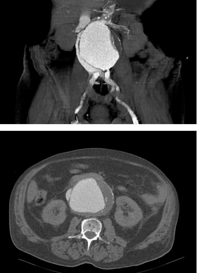
The chest x-ray showed a vascular redistribution pattern with small bilateral pleural effusion. A renal Doppler ultrasound showed normal-sized kidneys without signs of obstruction, a dilated right renal vein, and a large AAA. The CT scan with contrast showed an infrarenal, 10cm AAA with atherosclerosis and thrombosis, without retroperitoneal haematoma, as well as the flow of contrast to the vena cava and right renal vein during the arterial phase and the ACF (Figure 1).
With the diagnosis of ARF and CHF secondary to ACF, haemodialysis and surgery were performed, with control of the aneurysm neck, which required the ligation of left renal vein, the opening the aneurysmal sac (with the loss of 2 litres of blood), the closure of the ACF and placement of aorto-aortic bypass. The immediate postoperative diuresis was 70ml/h, but the patient developed refractory multiorgan failure and died three days after surgery.
The rupture of an aortic aneurysm and into other organs is uncommon. The inferior vena cava is the most common, followed by the iliac veins, the left renal vein and intestine.1,2-4 Over 80% of ACF cases are due to the rupture of an atherosclerotic aortic aneurysm (average size 11cm),2-5 with other causes being penetrating abdominal trauma; iatrogenia, mainly lumbar disc surgery or catheterisation; and occasionally others.1,3
Between 31% and 76% of the ACFs are detected during surgery after evacuating the clot from the aneurysm sac, which causes massive bleeding and/or a paradoxical pulmonary embolism.3,5-8 They are difficult to Identify because 20%-70% are associated with abdominal or retroperitoneal rupture where the characteristic signs (pulsatile abdominal mass, pain, signs of bleeding, abdominal murmur) are absent or may be easily misinterpreted. An aneurysmal thrombus in the fistula, arterial hypotension or compression of the inferior vena cava may mask the clinical signs of an ACF.1,3-5,7-9
The appearance of an ACF involves that aortic arterial blood is flowing to the vena cava, and therefore, arterial resistance is reduced, the renin-angiotensin-aldosterone and sympathetic systems are activated, and a hyperdynamic circulatory state is seen in 30%-50% of cases. If the ACF is large or the heart is unable to increase cardiac output, heart failure and refractory shock appear.1,6,8 In addition, ACF causes regional venous hypertension due to volume overload that manifests as oedema in the extremities or scrotum, haematuria, priapism, bleeding or dilated abdominal wall veins. The CVP may be low, normal or high, even very high, as in our patient, which should lead to suspecting the possible existence of ACF.3,7
ACF-associated renal manifestations are macroscopic or microscopic haematuria due to renal and/or bladder venous congestion (which is suggestive of ACF in AAA patients)3-5,7,10 and ARF, present in 7%-77% of cases and related to functional haemodynamic changes, renal venous hypertension and perfusion deficit.3,5,6 The immediate recovery of diuresis after surgical closure of the ACF and even the absence of renal lesions in the renal biopsy have been reported on many occasions.11,12
The diagnostic method of choice for ACF is CT, which shows early enhancement of the vena cava, isodense with the aneurysm and even the arteriovenous communication. Other findings include the inferior vena cava dilated or compressed by the aneurysm, retrograde opacification of the renal veins during the arterial phase, poor renal enhancement or an increase in size, and pelvic and retroperitoneal congestion.13,14
Post-operative-associated ACF mortality is 16%-72%, and is mainly related to incorrect diagnosis, hypotension, shock with anuria or ruptured aortic aneurysm.2-4,7-9
Specific treatment for ACF is transaortic closure and placement of a prosthetic graft, which improves heart and renal failure.3,5,7 A proposed alternative treatment to surgery is aortic stenting in the rupture of aortic aneurysms, with good results at centres with extensive experience.15
In summary, ACF is a rare complication of an aortic aneurysm, which requires high diagnostic suspicion because of its high mortality. The ACF may present with haematuria and/or oligoanuric acute renal failure requiring dialysis. Decreased renal perfusion and venous hypertension are the major pathophysiological mechanisms involved in the renal failure of these patients.
Figure 1. CT scan with contrast. Abdominal aorta aneurism and aortocaval fistula


