Las primeras biopsias realizadas, tanto a adultos como a niños, fueron quirúrgicas. Se realizaban a pacientes que se sometían a la decapsulación de los riñones con la intención de reducir la presión intrarrenal, generalmente en casos de síndrome nefrótico. En 1944, Nils Alwall inició la realización de biopsias renales percutáneas mediante aguja y aspiración en la Universidad de Lund (Suecia), aunque su experiencia se publicó en 1952. El primer artículo que tenía por tema la práctica de una biopsia renal percutánea fue escrito en 1950 por un médico cubano, Antonino Pérez Ara, y se publicó en una Revista local con escasa difusión. El primer trabajo que apareció en una Revista española (1953) sobre la práctica de las biopsias renales percutáneas no estaba firmado por ningún grupo español, sino por miembros del Hospital Calixto García, de la Universidad de La Habana, Cuba. El primer artículo publicado en España sobre el tema que nos ocupa vio la luz en 1958, hace ahora 50 años, en la Revista Clínica Española. Los dos primeros firmantes fueron Alfonso de la Peña Pineda y Vicente Gilsanz García, profesores de la Facultad de Medicina de Madrid. Posteriormente, la práctica de la biopsia renal percutánea se generalizó en los demás hospitales españoles.
The first renal biopsies, made as much in adults as in children, were surgical. They were made to patients who were under renal decapsulation with the intention to reduce the kidney pressure, especially in cases of nephrotic syndrome. In 1944, Nils Alwall initiated the accomplishment of percutaneous kidney biopsies by means of a needle and aspiration at the University of Lund (Sweden), although his experience was published in 1952. The first article that had by subject the practice of a percutaneous renal biopsy was written in 1950 by a Cuban doctor, Antonino Pérez Ara, and published in a local journal with little diffusion. The first work that appeared in a Spanish journal (1953) about the practice of the percutaneus renal biopsies was not signed by any Spanish group but by members of the Hospital “Calixto García” of the University of The Havana, Cuba. The first article published in Spain regarding to this subject, saw the light in 1958, now 50 years ago, in the Revista Clínica Española. The two first signers were Alfonso de la Peña Pineda and Vicente Gilsanz García, professors of the Medicine Faculty of Madrid. Later, the practice of the percutaneous renal biopsy became general in other Spanish hospitals.
INTRODUCTION
The practice of percutaneous renal biopsy was preceded by experience accumulated over three centuries based on macro and microscopic observations of renal tissue samples obtained from autopsies, the pioneers of which were Marcello Malpighi (1628-1694),1 Giovanni Battista Morgagni (1682-1771) and Marie François Xavier Bichat (1771-1802.) These techniques reached new heights during the 19th century with studies from Richard Bright (1789-1858)2 and Pierre Rayer (1793-1867)3 during the first third of this century and those of Friedrich Gustav Jacob Henle (1809-1885)4 and William Bowman (1816-1892)5 in the second third.
THE FIRST SURGICAL BIOPSIES IN ADULTS
According to Cameron and Hicks,6 the first surgical renal biopsies were performed on adult patients in New York by the gynaecologist and surgeon George Michael Edelbohls (1853- 1908.) The basic idea was that by making an incision in the renal capsule or performing a complete bilateral decapsulation, intrarenal pressure in cases of Bright’s disease or acute haemorrhagic nephritis could be reduced. Edelbohls published several articles on the surgical treatment of Bright’s disease. A book was published in 1904 reporting on an experiment he carried out on 72 patients, 16 of which experienced reduced symptoms and regained normal urine.
This text explains that in some cases of Bright’s disease the diagnosis was confirmed via a histological examination of the renal tissue.7 The first images of histological renal studies were published by Norman Gwin, Toronto, in 1923. The images show acute nephritis and amyloidosis.8 Renal capsulotomy continued to be performed throughout the first three decades of the 20th century, particularly in children.
THE FIRST SURGICAL BIOPSIES IN CHILDREN
As in the case of adults, the first surgical biopsies in children were performed on patients who underwent kidney decapsulation to treat nephropathy. In 1899, Ferguson published cases of two patients with Bright’s disease (age not given) who presented “chronic symptoms and had not responded to medication or test”.9
After decapsulating the kidneys, both patients improved in all their symptoms. Taking the opportunity of the surgical intervention, small fragments from the kidneys were taken to be examined in the microscope “showing that the diagnosis was correct.”9
A few years later, in 1904, Graham, from Philadelphia, published the case of a young girl aged 26 months with generalised anasarca and massive albuminuria. Since the child did not respond to conventional treatment and death was imminent, decapsulation of the kidneys was performed.10 A small portion of the right kidney was extracted and submitted to histological examination. Microscopic examination showed that some of the glomeruli were abnormally cellular. Other glomeruli were quite retracted and the glomerular space was filled with a slightly acidophilic and granular substance. The epithelium lining of the “uriniferous tubules” was inflamed, dark or granular and, in some places, actively scaling.11
In Europe, the first open renal biopsies were performed in children from 1917 in the Royal Hospital for Sick Children in Glasgow. As previously indicated, these were performed during interventions for renal decapsulation in cases of nephrotic syndrome. The results of these first biopsies, 23 to be exact, were published by Campbell.12,13 It is to be noted that these children, who had large oedemas and were prone to suffering infections, showed a 50% recovery rate.13 In England, renal biopsies for decapsulation began to be performed in the Royal Liverpool Children’s Hospital from 1923 by Norman Capon.14 Incidentally, renal function was tested by determining the excretion of phenolsulphtalein during the first two hours following the intramuscular injection of this substance.6
THE FIRST PERCUTANEOUS BIOPSIES
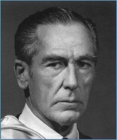
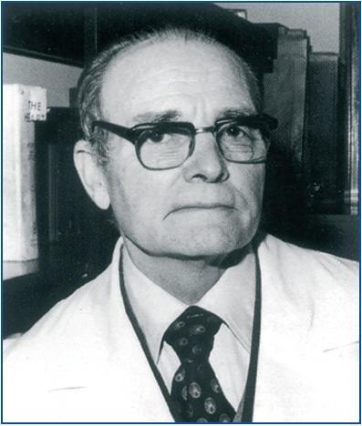
In 1939, Poul Iversen and Kaj Roholm, from Copenhagen, described a percutaneous liver biopsy using a 1mm needle and a syringe for aspiration.15 In 1944, following the previous experiments of Iversen and Roholm, Nils Alwall (1904-1986) began to perform percutaneous renal biopsies using a needle and aspiration for the first time in the University of Lund (Sweden), however the experiment was not published until 1952.16 Asimple radiograph and retrograde pyelogram were used to locate the kidney. Dr Alwall is known in the history of nephrology for being one of the pioneers in practicing haemodialysis (figure 1.)
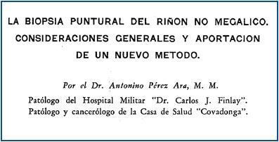
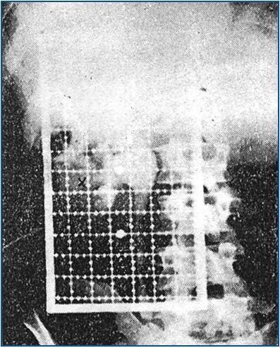
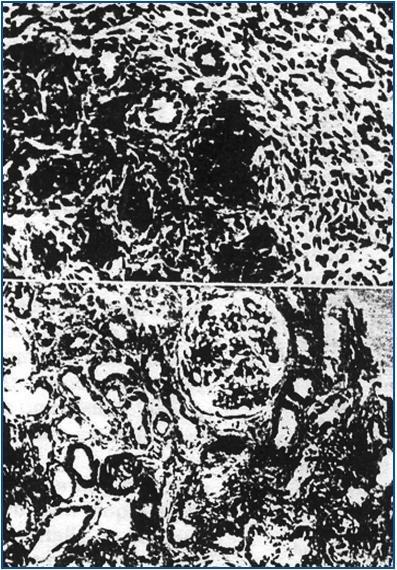
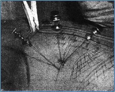
The first article published on the practice of percutaneous renal biopsy was written by a Cuban doctor, Antonino Pérez Ara, in 1950. It appeared in a local journal with small circulation, two years before the previously mentioned article by Nils Alwall17 (figure 2.) Therefore, it is often forgotten that this was a pioneering article. Dr Pérez Ara was a pathologist at the Dr Carlos J Finlay Military Hospital and a pathologist and cancer specialist at the Covadonga Clinic. Having no knowledge of the studies of Nils Alwall, he began to develop his technique “during the last months of 1948, practicing it on eight patients.” The kidney was located using a pyelogram performed via an endovenous injection of “20cc of Diodrast” with the help of “a grid to help select the best site for the puncture on the subject’s skin” (figure 3.) “A special type of trocar was used which the author called a nefrobiótomo (nephro-bioptome), which is simply the Herrera-Pardo biopsy needle which had been specially modified for this new application”.17 The patient was positioned in the prone position and either one of the kidneys was punctured.
The study of Pérez-Ara was unknown to the abovementioned internist Poul Iversen and Claus Brun (one of the first nephrologists) from Copenhagen, who began to use a technique which was almost identical to that used for aspiration biopsy used in cases of liver biopsies.18 A biopsy was taken of the right kidney, with the patient sat down, using an Iversen and Roholm needle after the organ was located using a radiograph. Suitable fragments were obtained on 42 occasions (53%) in 60 patients who underwent the procedure a total of 80 times (figure 4.) In this instance, renal function studies included determining the urea, creatinine and paraaminohippurate clearance, as well as the Addis count. Their experience was published in 1951.18 Claus Brun was the second President of the International Society of Nephrology (1963-1966), after Jean Hamburger.6
Following the Danish publication, several groups in different countries all over the world began to perform percutaneous renal biopsies. Thus, details were published of experiences carried out in 1952 by the Italians Aldo Torsoli and Enrico Fiaschi6 and in 1953 by the Maurice Payet group in Dakar (Senegal),19 the Alvin Parrish and John Howe group in Washington20 and Grenwald et al. in Brooklyn.21
However, soon after fatal cases attributed to renal biopsy were published,22 as had previously occurred with one of Alwall’s patients.16
Robert Kark and Robert Muehrcke from the University of Illinois College of Medicine, Chicago, stated that the practice of biopsies on patients who were sat down was an unsatisfactory procedure both for the person performing the procedure and the patient. Thus, suitable samples were obtained in 48 of the first 50 attempts using the Vim-Silverman needle in the prone position.23
By the end of the 1950s, in addition to the previously mentioned articles, percutaneous renal biopsies had also been published in Switzerland, Poland, Hungary, the United Kingdom, France, Canada, Germany, India, Sweden, Argentina, Japan, Spain, the Czech Republic, Russia, Romania, Finland, Uruguay, Brazil, Portugal and Austria, as well as the incorporation of new groups in the United States.6
THE FIRST PERCUTANEOUS BIOPSIES IN SPAIN
The first article published in Spain on the practice of percutaneous renal biopsies was not written by a Spanish group. Drs Pardo, Cárdenas and Masó from the Hospital Calixto García in Havana published an article in the journal Revista Clínica Española in 1953, describing their experience in this area.24 The point to be punctured was located using retro-pneumoperitoneum with oxygen. On arriving “at the Xray department, a metal squared grid was positioned on the lumbar region. A plate was then taken in anteroposterior position, observing on the negative the relationship between the renal shadow and the metal mesh and the selected point was immediately marked on the skin.” The biopsies were performed in prone position with the help of a Silverman trocar. Victoriano Pardo et al. obtained “55 satisfactory samples from 80 patients and a total of 90 attempts.”
From a personal communication belonging to Dr Luis Hernando Avendaño, it is known that from mid-50s the Jiménez Díaz Foundation was performing surgical renal biopsies, “which were studied on Tuesdays by Mr Carlos Jiménez Díaz, Morales and Oliva-Horacio.”
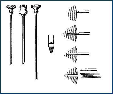
The first article on this issue in Spain was published in the journal Revista Clínica Española in 1958, 50 years ago.25 The first two signatories of the article were Alfonso de la Peña Pineda (1904-1971) (figure 5) and Vicente Gilsanz García (1911-1992) (figure 6) who, at the time, were Professors of Urology and Pathology and Clinical Medicine at the Faculty of Medicine of Madrid, respectively. The patient was in prone position and the Franklin-Vim Silverman needle was used to puncture either of the two kidneys (figure 7.) The authors showed that they had vast experience, performing a total of 94 biopsies, which took place without any complications. The most frequent diagnoses were renal sclerosis, chronic nephritis, pyelonephritis, Wilson’s disease and nephrosis. The high number of patients with Wilson’s disease who underwent biopsies was due to “attempts to meticulously complete a study on Wilson’s disease. It was always discussed whether the aminoaciduria and copper levels in patients with hepatolenticular degeneration was due to functional or organ disease, since no histological alterations in the kidney had been confirmed until now.” Months later, the same group from the Faculty of Medicine of Madrid published an extended experiment with 112 biopsies in La Presse Medicale26. In this case, the results related to patients with Wilson’s disease were not mentioned either.
In the same year, 1958, Gerardo del Río wrote a chapter on renal biopsy in the well-known book on Internal Medicine by Agustín Pedro Pons.27 The author described the technique of Kark y Muehrcke28 and, given the references to several histological images in the book, it is possible that they had also begun performing renal biopsies at that time in Barcelona. Pedro Pons and Del Río published an article on lupus nephritis which included the content of a paper presented at the 3rd Spanish Congress on Internal Medicine (III Congreso Nacional de Medicina Interna), which was held in Madrid in June 1958. The article states: “The anatomical pathology of this disease is now well known, and the accessibility of renal parenchyma in puncture biopsies gives it particular importance.”
The results obtained in biopsies performed on patients with Wilson’s disease were published in Spanish in 195929 and in English the following year.30 “The results of 17 biopsies carried out on five patients with Wilson’s disease showed that those patients suffered no morphological modifications in the nephron.” In 1962, Gerardo del Río wrote about his experiment on lupus nephritis. “It was possible to perform a histological check in 17 cases, an autopsy check in nine and a biopsy in eight. For the percutaneous renal biopsy the Kark and Muhercke technique was used, with a Vim-Silverman needle”.31 This year, the same author published his experiment on Schönlein- Henoch nephropathy.32
In the same year, 1962, Gilsanz et al. revised their experiment based on an article published in the records of the Faculty of Medicine, Madrid.33 At this point “they had performed 348 biopsies on 276 patients.” In this article, they commented on some patients in more detail. For example, acute haemorrhagic nephritis in a case of an addisonian, a rarity since “Dr Gregorio Marañón had not ever seen this in his vast experience of this disease.” Other diagnoses cited were evolving pyelonephritis, membranous glomerulitis (sic) and chronic lobular glomerulitis.
In 1968, the Departments of Nephrology and Pathological Anatomy at the Jiménez Díaz Foundation in Madrid published an overview of percutaneous renal biopsy in the journal Revista Clínica Española.34 The number of patients who underwent biopsy was not specified, however, two clinical cases were mentioned: the first was a patient with renal amyloidosis, and the second was a patient with subacute glomerulonephritis (figure 8.) In 1973, the number of biopsies performed in the Clinical Hospital of Madrid amounted to 1650.35 As the end of the history of nephrology is reached, there is one point that was undoubtedly significant and needs to be highlighted. None of the mentioned articles, which were written by Spanish authors, cited the other Spanish authors who also worked on the issue. Awatchful reader will note that the text refers to the three main hospitals and medical science centres in Spain during the second third of the 20th century, which also included three great and unrivalled figures in the history of Internal Medicine in Spain. These include, indeed, Dr Gregorio Marañón (1887-1960), (Clinical Hospital of Madrid), Dr Agustín Pedro Pons (1898-1971) (Clinical Hospital of Barcelona) and Dr Carlos Jiménez Díaz (1898-1967) (Jiménez Díaz Foundation.) Although it seems unlikely from these doctors, it is probable that there was a “scientific struggle” between the three centres; therefore, they all may preferred to ignore one another.
Figure 1.
Figure 2.
Figure 3.
Figure 4.
Figure 5.
Figure 6.
Figure 7.
Figure 8.


