array:21 [
  "pii" => "X2013251410050418"
  "issn" => "20132514"
  "doi" => "10.3265/Nefrologia.pre2010.May.10452"
  "estado" => "S300"
  "fechaPublicacion" => "2010-07-01"
  "documento" => "article"
  "licencia" => "http://www.elsevier.com/open-access/userlicense/1.0/"
  "subdocumento" => "fla"
  "cita" => "Nefrologia (English Version). 2010;30:463-6"
  "abierto" => array:3 [
    "ES" => true
    "ES2" => true
    "LATM" => true
  ]
  "gratuito" => true
  "lecturas" => array:2 [
    "total" => 11659
    "formatos" => array:3 [
      "EPUB" => 289
      "HTML" => 10650
      "PDF" => 720
    ]
  ]
  "Traduccion" => array:1 [
    "es" => array:17 [
      "pii" => "X0211699510050410"
      "issn" => "02116995"
      "doi" => "10.3265/Nefrologia.pre2010.May.10452"
      "estado" => "S300"
      "fechaPublicacion" => "2010-07-01"
      "documento" => "article"
      "licencia" => "http://www.elsevier.com/open-access/userlicense/1.0/"
      "subdocumento" => "fla"
      "cita" => "Nefrologia. 2010;30:463-6"
      "abierto" => array:3 [
        "ES" => true
        "ES2" => true
        "LATM" => true
      ]
      "gratuito" => true
      "lecturas" => array:2 [
        "total" => 36587
        "formatos" => array:3 [
          "EPUB" => 338
          "HTML" => 35080
          "PDF" => 1169
        ]
      ]
      "es" => array:12 [
        "idiomaDefecto" => true
        "titulo" => "Catéter en vena cava superior para hemodiálisis entre los últimos recursos en hemitórax superior"
        "tienePdf" => "es"
        "tieneTextoCompleto" => "es"
        "tieneResumen" => array:2 [
          0 => "es"
          1 => "en"
        ]
        "paginas" => array:1 [
          0 => array:2 [
            "paginaInicial" => "463"
            "paginaFinal" => "466"
          ]
        ]
        "titulosAlternativos" => array:1 [
          "en" => array:1 [
            "titulo" => "Catheter in the superior vena cava for hemodialysis as a last resort in superior hemithorax"
          ]
        ]
        "contieneResumen" => array:2 [
          "es" => true
          "en" => true
        ]
        "contieneTextoCompleto" => array:1 [
          "es" => true
        ]
        "contienePdf" => array:1 [
          "es" => true
        ]
        "resumenGrafico" => array:2 [
          "original" => 0
          "multimedia" => array:8 [
            "identificador" => "fig1"
            "etiqueta" => "Fig. 1"
            "tipo" => "MULTIMEDIAFIGURA"
            "mostrarFloat" => true
            "mostrarDisplay" => false
            "copyright" => "Elsevier España"
            "figura" => array:1 [
              0 => array:4 [
                "imagen" => "10452_18030_4264_es_10452figura1.jpg"
                "Alto" => 273
                "Ancho" => 394
                "Tamanyo" => 49411
              ]
            ]
            "descripcion" => array:1 [
              "es" => "Paciente D en segundo día postquirurgico."
            ]
          ]
        ]
        "autores" => array:1 [
          0 => array:2 [
            "autoresLista" => "César Augusto Restrepo Valencia, C.M. Buritica Barragán, A. Arango"
            "autores" => array:3 [
              0 => array:2 [
                "nombre" => "César Augusto"
                "apellidos" => "Restrepo Valencia"
              ]
              1 => array:2 [
                "Iniciales" => "C.M."
                "apellidos" => "Buritica Barragán"
              ]
              2 => array:2 [
                "Iniciales" => "A."
                "apellidos" => "Arango"
              ]
            ]
          ]
        ]
      ]
      "idiomaDefecto" => "es"
      "Traduccion" => array:1 [
        "en" => array:9 [
          "pii" => "X2013251410050418"
          "doi" => "10.3265/Nefrologia.pre2010.May.10452"
          "estado" => "S300"
          "subdocumento" => ""
          "abierto" => array:3 [
            "ES" => true
            "ES2" => true
            "LATM" => true
          ]
          "gratuito" => true
          "lecturas" => array:1 [
            "total" => 0
          ]
          "idiomaDefecto" => "en"
          "EPUB" => "https://multimedia.elsevier.es/PublicationsMultimediaV1/item/epub/X2013251410050418?idApp=UINPBA000064"
        ]
      ]
      "EPUB" => "https://multimedia.elsevier.es/PublicationsMultimediaV1/item/epub/X0211699510050410?idApp=UINPBA000064"
      "url" => "/02116995/0000003000000004/v0_201502091427/X0211699510050410/v0_201502091427/es/main.assets"
    ]
  ]
  "itemSiguiente" => array:17 [
    "pii" => "X201325141005040X"
    "issn" => "20132514"
    "doi" => "10.3265/Nefrologia.pre2010.May.10390"
    "estado" => "S300"
    "fechaPublicacion" => "2010-07-01"
    "documento" => "article"
    "licencia" => "http://www.elsevier.com/open-access/userlicense/1.0/"
    "subdocumento" => "fla"
    "cita" => "Nefrologia (English Version). 2010;30:467-72"
    "abierto" => array:3 [
      "ES" => true
      "ES2" => true
      "LATM" => true
    ]
    "gratuito" => true
    "lecturas" => array:2 [
      "total" => 6216
      "formatos" => array:3 [
        "EPUB" => 280
        "HTML" => 5303
        "PDF" => 633
      ]
    ]
    "en" => array:12 [
      "idiomaDefecto" => true
      "titulo" => "Sjögren's syndrome and acute renal failure after oral surgery"
      "tienePdf" => "en"
      "tieneTextoCompleto" => "en"
      "tieneResumen" => array:2 [
        0 => "es"
        1 => "en"
      ]
      "paginas" => array:1 [
        0 => array:2 [
          "paginaInicial" => "467"
          "paginaFinal" => "472"
        ]
      ]
      "titulosAlternativos" => array:1 [
        "es" => array:1 [
          "titulo" => "Síndrome de Sjögren y fracaso renal agudo tras cirugía oral"
        ]
      ]
      "contieneResumen" => array:2 [
        "es" => true
        "en" => true
      ]
      "contieneTextoCompleto" => array:1 [
        "en" => true
      ]
      "contienePdf" => array:1 [
        "en" => true
      ]
      "resumenGrafico" => array:2 [
        "original" => 0
        "multimedia" => array:8 [
          "identificador" => "fig1"
          "etiqueta" => "Fig. 1"
          "tipo" => "MULTIMEDIAFIGURA"
          "mostrarFloat" => true
          "mostrarDisplay" => false
          "copyright" => "Elsevier España"
          "figura" => array:1 [
            0 => array:4 [
              "imagen" => "10390_108_7203_en_10390_figura_1.jpg"
              "Alto" => 480
              "Ancho" => 640
              "Tamanyo" => 205095
            ]
          ]
          "descripcion" => array:1 [
            "en" => "PAS. Massive glomerular intracapillary thrombi"
          ]
        ]
      ]
      "autores" => array:1 [
        0 => array:2 [
          "autoresLista" => "R. Toledo Rojas, I. García, A. Torres, M.A. Frutos, G. Martín-Reyes, C. Jironda, R. Franquelo, M. León, D. Hernández"
          "autores" => array:9 [
            0 => array:2 [
              "Iniciales" => "R."
              "apellidos" => "Toledo Rojas"
            ]
            1 => array:2 [
              "Iniciales" => "I."
              "apellidos" => "García"
            ]
            2 => array:2 [
              "Iniciales" => "A."
              "apellidos" => "Torres"
            ]
            3 => array:2 [
              "Iniciales" => "M.A."
              "apellidos" => "Frutos"
            ]
            4 => array:2 [
              "Iniciales" => "G."
              "apellidos" => "Martín-Reyes"
            ]
            5 => array:2 [
              "Iniciales" => "C."
              "apellidos" => "Jironda"
            ]
            6 => array:2 [
              "Iniciales" => "R."
              "apellidos" => "Franquelo"
            ]
            7 => array:2 [
              "Iniciales" => "M."
              "apellidos" => "León"
            ]
            8 => array:2 [
              "Iniciales" => "D."
              "apellidos" => "Hernández"
            ]
          ]
        ]
      ]
    ]
    "idiomaDefecto" => "en"
    "Traduccion" => array:1 [
      "es" => array:9 [
        "pii" => "X0211699510050402"
        "doi" => "10.3265/Nefrologia.pre2010.May.10390"
        "estado" => "S300"
        "subdocumento" => ""
        "abierto" => array:3 [
          "ES" => true
          "ES2" => true
          "LATM" => true
        ]
        "gratuito" => true
        "lecturas" => array:1 [
          "total" => 0
        ]
        "idiomaDefecto" => "es"
        "EPUB" => "https://multimedia.elsevier.es/PublicationsMultimediaV1/item/epub/X0211699510050402?idApp=UINPBA000064"
      ]
    ]
    "EPUB" => "https://multimedia.elsevier.es/PublicationsMultimediaV1/item/epub/X201325141005040X?idApp=UINPBA000064"
    "url" => "/20132514/0000003000000004/v0_201502091652/X201325141005040X/v0_201502091652/en/main.assets"
  ]
  "itemAnterior" => array:17 [
    "pii" => "X2013251410050426"
    "issn" => "20132514"
    "doi" => "10.3265/Nefrologia.pre2010.Mar.10321"
    "estado" => "S300"
    "fechaPublicacion" => "2010-07-01"
    "documento" => "article"
    "licencia" => "http://www.elsevier.com/open-access/userlicense/1.0/"
    "subdocumento" => "fla"
    "cita" => "Nefrologia (English Version). 2010;30:458-62"
    "abierto" => array:3 [
      "ES" => true
      "ES2" => true
      "LATM" => true
    ]
    "gratuito" => true
    "lecturas" => array:2 [
      "total" => 6012
      "formatos" => array:3 [
        "EPUB" => 273
        "HTML" => 5137
        "PDF" => 602
      ]
    ]
    "en" => array:12 [
      "idiomaDefecto" => true
      "titulo" => "Differences between the CKD-EPI and the MDRD equations when estimating the glomerular filtration rate in hypertensive patients"
      "tienePdf" => "en"
      "tieneTextoCompleto" => "en"
      "tieneResumen" => array:2 [
        0 => "es"
        1 => "en"
      ]
      "paginas" => array:1 [
        0 => array:2 [
          "paginaInicial" => "458"
          "paginaFinal" => "462"
        ]
      ]
      "titulosAlternativos" => array:1 [
        "es" => array:1 [
          "titulo" => "Diferencias de la ecuación CKD-EPI con la de MDRD para la estimación del filtrado glomerular en pacientes hipertensos"
        ]
      ]
      "contieneResumen" => array:2 [
        "es" => true
        "en" => true
      ]
      "contieneTextoCompleto" => array:1 [
        "en" => true
      ]
      "contienePdf" => array:1 [
        "en" => true
      ]
      "resumenGrafico" => array:2 [
        "original" => 0
        "multimedia" => array:8 [
          "identificador" => "fig1"
          "etiqueta" => "Fig. 1"
          "tipo" => "MULTIMEDIAFIGURA"
          "mostrarFloat" => true
          "mostrarDisplay" => false
          "copyright" => "Elsevier España"
          "figura" => array:1 [
            0 => array:4 [
              "imagen" => "10321_108_7349_en_10321_f1.jpg"
              "Alto" => 426
              "Ancho" => 510
              "Tamanyo" => 27248
            ]
          ]
          "descripcion" => array:1 [
            "en" => "Bland-Atmann graph"
          ]
        ]
      ]
      "autores" => array:1 [
        0 => array:2 [
          "autoresLista" => "M.A. Gómez Marcos, E. Rodríguez Hernández, E. Rodríguez Sánchez, J.I. Recio Rodríguez, C. Martín, C. Martín Cantera, Rafael Ramos Blanes, R. Ramos Blanes, Luis García Ortiz, L. García Ortiz"
          "autores" => array:10 [
            0 => array:2 [
              "Iniciales" => "M.A."
              "apellidos" => "Gómez Marcos"
            ]
            1 => array:2 [
              "Iniciales" => "E."
              "apellidos" => "Rodríguez Hernández"
            ]
            2 => array:2 [
              "Iniciales" => "E."
              "apellidos" => "Rodríguez Sánchez"
            ]
            3 => array:2 [
              "Iniciales" => "J.I."
              "apellidos" => "Recio Rodríguez"
            ]
            4 => array:2 [
              "Iniciales" => "C."
              "apellidos" => "Martín"
            ]
            5 => array:2 [
              "Iniciales" => "C."
              "apellidos" => "Martín Cantera"
            ]
            6 => array:2 [
              "nombre" => "Rafael"
              "apellidos" => "Ramos Blanes"
            ]
            7 => array:2 [
              "Iniciales" => "R."
              "apellidos" => "Ramos Blanes"
            ]
            8 => array:2 [
              "nombre" => "Luis"
              "apellidos" => "García Ortiz"
            ]
            9 => array:2 [
              "Iniciales" => "L."
              "apellidos" => "García Ortiz"
            ]
          ]
        ]
      ]
    ]
    "idiomaDefecto" => "en"
    "Traduccion" => array:1 [
      "es" => array:9 [
        "pii" => "X0211699510050429"
        "doi" => "10.3265/Nefrologia.pre2010.Mar.10321"
        "estado" => "S300"
        "subdocumento" => ""
        "abierto" => array:3 [
          "ES" => true
          "ES2" => true
          "LATM" => true
        ]
        "gratuito" => true
        "lecturas" => array:1 [
          "total" => 0
        ]
        "idiomaDefecto" => "es"
        "EPUB" => "https://multimedia.elsevier.es/PublicationsMultimediaV1/item/epub/X0211699510050429?idApp=UINPBA000064"
      ]
    ]
    "EPUB" => "https://multimedia.elsevier.es/PublicationsMultimediaV1/item/epub/X2013251410050426?idApp=UINPBA000064"
    "url" => "/20132514/0000003000000004/v0_201502091652/X2013251410050426/v0_201502091652/en/main.assets"
  ]
  "en" => array:15 [
    "idiomaDefecto" => true
    "titulo" => "Catheter in the superior vena cava for hemodialysis as a last resort in superior hemithorax"
    "tieneTextoCompleto" => true
    "paginas" => array:1 [
      0 => array:2 [
        "paginaInicial" => "463"
        "paginaFinal" => "466"
      ]
    ]
    "autores" => array:1 [
      0 => array:3 [
        "autoresLista" => "César Augusto Restrepo Valencia, C.M. Buritica Barragán, A. Arango"
        "autores" => array:3 [
          0 => array:4 [
            "nombre" => "César Augusto"
            "apellidos" => "Restrepo Valencia"
            "email" => array:1 [
              0 => "caugustorv@une.net.co"
            ]
            "referencia" => array:1 [
              0 => array:2 [
                "etiqueta" => "<span class="elsevierStyleSup">a</span>"
                "identificador" => "affa"
              ]
            ]
          ]
          1 => array:3 [
            "Iniciales" => "C.M."
            "apellidos" => "Buritica Barrag&#225;n"
            "referencia" => array:1 [
              0 => array:2 [
                "etiqueta" => "<span class="elsevierStyleSup">b</span>"
                "identificador" => "affb"
              ]
            ]
          ]
          2 => array:3 [
            "Iniciales" => "A."
            "apellidos" => "Arango"
            "referencia" => array:1 [
              0 => array:2 [
                "etiqueta" => "<span class="elsevierStyleSup">c</span>"
                "identificador" => "affc"
              ]
            ]
          ]
        ]
        "afiliaciones" => array:3 [
          0 => array:3 [
            "entidad" => "Profesor Asociado, Universidad de Caldas,   Colombia, "
            "etiqueta" => "<span class="elsevierStyleSup">a</span>"
            "identificador" => "affa"
          ]
          1 => array:3 [
            "entidad" => "Médico RTS Ltda., Sucursal Caldas,   Colombia, "
            "etiqueta" => "<span class="elsevierStyleSup">b</span>"
            "identificador" => "affb"
          ]
          2 => array:3 [
            "entidad" => "Cirujano de Tórax, Universidad de Caldas,   Colombia, "
            "etiqueta" => "<span class="elsevierStyleSup">c</span>"
            "identificador" => "affc"
          ]
        ]
      ]
    ]
    "titulosAlternativos" => array:1 [
      "es" => array:1 [
        "titulo" => "Cat&#233;ter en vena cava superior para hemodi&#225;lisis entre los &#250;ltimos recursos en hemit&#243;rax superior"
      ]
    ]
    "resumenGrafico" => array:2 [
      "original" => 0
      "multimedia" => array:8 [
        "identificador" => "fig1"
        "etiqueta" => "Tab.  1"
        "tipo" => "MULTIMEDIAFIGURA"
        "mostrarFloat" => true
        "mostrarDisplay" => false
        "copyright" => "Elsevier Espa&#241;a"
        "figura" => array:1 [
          0 => array:4 [
            "imagen" => "10452_108_7891_en_10452_t1.jpg"
            "Alto" => 518
            "Ancho" => 849
            "Tamanyo" => 63101
          ]
        ]
        "descripcion" => array:1 [
          "en" => "Patient characteristics and outcomes"
        ]
      ]
    ]
    "textoCompleto" => "<p class="elsevierStylePara"><span class="elsevierStyleBold">INTRODUCTION </span></p><p class="elsevierStylePara">In patients with chronic kidney disease &#40;CKD&#41; on haemodialysis therapy&#44; vascular access for the procedure is one of the major problems faced by nephrologists on a daily basis&#46; Arteriovenous &#40;A-V&#41; fistulas with native vessels or grafts are ideal forms of access due to their long life and low rate of complications during use&#46; For patients in whom an A-V fistula cannot be used&#44; central venous catheters must be used&#44; although they frequently cause thrombosis of the vessels where they are placed and they exhaust the classic vessels &#40;internal jugular&#44; subclavian&#41; and non-classic vessels &#40;axillary and innominate&#41; in the upper hemithorax&#46;</p><p class="elsevierStylePara">Before using infradiaphragmatic vessels or&#44; for those patients where the use of those vessels is not possible&#44; there are two final alternatives for placing central lines&#58; intracardiac access &#40;right atrium&#41; and direct puncture of the superior vena cava&#46; In this report we present the placement of 4 catheters for haemodialysis in the superior vena cava using parasternal access&#46;</p><p class="elsevierStylePara">&#160;</p><p class="elsevierStylePara"><span class="elsevierStyleBold">CASE REPORTS</span></p><p class="elsevierStylePara">Four patients who had previously had catheters placed in various supradiaphragmatic veins with ultrasound-documented thrombosis of the internal jugular&#44; subclavian&#44; axillary&#44; and innominate veins&#44; who were either not candidates for or refused peritoneal dialysis&#44; were selected for placement of a catheter in the superior vena cava&#46; All patients gave written consent for the procedure&#46; Exclusion criteria were age below 18&#44; coagulation test &#40;PTT and PT&#41; abnormalities&#44; thrombocytopoenia &#40;platelet count less than 50&#44;000 platelets&#41; and refusal to participate&#46; Patient characteristics and outcomes are shown in table 1&#46;</p><p class="elsevierStylePara">The surgical technique employed was as follows&#58; 1&#41; conventional preparation for surgery under general anaesthesia&#44; 2&#41; right anterior mediastinotomy&#44; with incision through the third intercostal space &#40;horizontally&#41; until resection of the chondrosternal junction&#44; 3&#41; ligation of the mammary vessels&#44; 4&#41; extrapleural approach to the superior vena cava&#44; 5&#41; creation of the subcutaneous tunnel and tunnelling of the catheter in the anterior thoracic wall with the catheter exit site at the midclavicular line of the fifth intercostal space&#44; 6&#41; under direct vision and after purse-string suture with 3-0 prolene&#44; the superior vena cava is punctured and the haemodialysis catheter &#40;indwelling type&#41; is placed&#44; directing the tip of the catheter downward and closing the purse-string suture&#44; 7&#41; the catheters are checked for patency and heparinised&#44; and 8&#41; mediastinotomy is closed in layers&#46;</p><p class="elsevierStylePara">This procedure was carried out successfully in 4 patients&#44; and the following complications were attributable to it&#58; three haemothorax episodes&#44; one of which was massive&#44; for which a thoracotomy was performed on each patient and a chest tube was placed for an average of 5 days&#59; the patient with massive haemothorax required transfusion of 5 units of red blood cells&#44; mediastinostomy&#44; ligation of the bleeding vessels&#44; and thoracotomy with chest tube for 7 days &#40;figures 1 and 2&#41;&#46;</p><p class="elsevierStylePara">Subsequently&#44; the patients were taken for chronic haemodialysis and progressed satisfactorily&#44; without complications attributable to the procedure&#46; Their last average Kt&#47;V was 1&#46;45 and one patient completed 36 months of using the catheter &#40;figure 3&#41;&#46;</p><p class="elsevierStylePara">&#160;</p><p class="elsevierStylePara"><span class="elsevierStyleBold">DISCUSSION</span></p><p class="elsevierStylePara">As the population of CKD patients on haemodialysis therapy ages&#44; it becomes increasingly difficult to obtain satisfactory access through which to provide their therapy&#46; A-V fistulas have the tremendous advantage of permitting multiple punctures over a long period of time&#44; but in a group of patients&#44; mainly diabetics&#44; it becomes impossible to place them&#46; The same is true with A-V prosthesis placement&#46; In this group of patients&#44; the use of either temporary or indwelling central venous catheters&#44; inserted through various sites&#44; becomes necessary&#46;</p><p class="elsevierStylePara">The internal jugular vein approach is the most commonly used due to its easy puncture and the low rate of complications&#46;<span class="elsevierStyleSup">1</span> The subclavian route is not recommended&#44; since it results in high stenosis and thrombosis rates&#44; which subsequently prevent the use of the upper extremity for the creation of A-V fistulas&#46;<span class="elsevierStyleSup">2</span> The axillary and innominate veins can also be used for the placement of central catheters&#44; but they require medical personnel familiar with those puncture procedures in order to be utilized&#46;<span class="elsevierStyleSup">3&#44;4</span> The infradiaphragmatic approach offers several routes&#58; the femoral&#44; with easy access but with the disadvantage of a high thrombosis and infection rates&#59;<span class="elsevierStyleSup">5</span> on the other hand&#44; the transhepatic and translumbar routes<span class="elsevierStyleSup">6&#44;7</span> are technically more difficult&#46; At the supradiaphragmatic level&#44; two final approaches allow for catheter implantation&#58; the intracardiac and the right parasternal routes&#44; each of which requires the use of general anaesthesia and anterior thoracotomy to access the puncture site&#46; In the intracardiac access the right atrium is punctured&#44; with later placement and tunneling of the indwelling catheter&#46;<span class="elsevierStyleSup">8</span></p><p class="elsevierStylePara">Right parasternal access was first described in the year 2002 by Archundia et al&#44;<span class="elsevierStyleSup">9</span>&#160;who performed it on a patient who had exhausted all supra- and infradiaphragmatic vascular access points&#46; In this technical report&#44; 3 more patients are being reported&#44; without establishing its long-term outcomes&#46;</p><p class="elsevierStylePara">The lack of literature indicating the use of this route after its initial introduction is particularly curious&#46;</p><p class="elsevierStylePara">In our area&#44; about 25&#37; of patients on haemodialysis therapy require the use of indwelling catheters for haemodialysis&#44; which has forced us to employ the vast majority of known routes for both supra- and infradiaphragmatic catheter&#44; including femoral&#44; iliac&#44; and translumbar catheters&#46; Nephrologists perform the aforementioned percutaneous procedures&#44; the majority with ultrasound guidance&#44; while fluoroscopy or computed axial tomography is used only for translumbar placement&#46; When it comes to catheters such as those presented in this case&#44; vascular or thoracic surgeons must be involved&#46; Otherwise&#44; such placement would be impossible&#44; since this medical group possesses the requisite skills and knowledge of intrathoracic anatomy&#46;</p><p class="elsevierStylePara">Based on our experience&#44; it has been possible to achieve satisfactory placement of 4 catheters in the superior vena cava for 36 months&#46; It is important to note that surgical complications are common&#44; despite the experience of the surgical team with thoracic surgeries&#46; Our recommendation is to always rely on experienced surgeons and to be alert to the occurrence of complications in order to resolve them quickly and avoid fatal consequences&#46;</p><p class="elsevierStylePara"><a href="grande&#47;10452&#95;108&#95;7891&#95;en&#95;10452&#95;t1&#46;jpg" class="elsevierStyleCrossRefs"><img src="10452_108_7891_en_10452_t1.jpg" alt="Patient characteristics and outcomes"></img></a></p><p class="elsevierStylePara">Table 1&#46; Patient characteristics and outcomes</p><p class="elsevierStylePara"><a href="grande&#47;10452&#95;108&#95;7892&#95;en&#95;10452&#95;f1&#46;jpg" class="elsevierStyleCrossRefs"><img src="10452_108_7892_en_10452_f1.jpg" alt="Patient D on second post-operative day&#46;"></img></a></p><p class="elsevierStylePara">Figure 1&#46; Patient D on second post-operative day&#46;</p><p class="elsevierStylePara"><a href="grande&#47;10452&#95;108&#95;7893&#95;en&#95;10452&#95;f2&#46;jpg" class="elsevierStyleCrossRefs"><img src="10452_108_7893_en_10452_f2.jpg" alt="PA chest x-ray of patient D&#46;"></img></a></p><p class="elsevierStylePara">Figure 2&#46; PA chest x-ray of patient D&#46;</p><p class="elsevierStylePara"><a href="grande&#47;10452&#95;108&#95;7894&#95;en&#95;10452&#95;f3&#46;jpg" class="elsevierStyleCrossRefs"><img src="10452_108_7894_en_10452_f3.jpg" alt="Patient 36 months after catheter placement&#46;"></img></a></p><p class="elsevierStylePara">Figure 3&#46; Patient 36 months after catheter placement&#46;</p>"
    "pdfFichero" => "P1-E501-S2445-A10452-EN.pdf"
    "tienePdf" => true
    "PalabrasClave" => array:2 [
      "es" => array:4 [
        0 => array:4 [
          "clase" => "keyword"
          "titulo" => "Palabras clave"
          "identificador" => "xpalclavsec441783"
          "palabras" => array:1 [
            0 => "Accesos vasculares"
          ]
        ]
        1 => array:4 [
          "clase" => "keyword"
          "titulo" => "Palabras clave"
          "identificador" => "xpalclavsec441785"
          "palabras" => array:1 [
            0 => "Vena cava superior"
          ]
        ]
        2 => array:4 [
          "clase" => "keyword"
          "titulo" => "Palabras clave"
          "identificador" => "xpalclavsec441787"
          "palabras" => array:1 [
            0 => "Hemodi&#225;lisis"
          ]
        ]
        3 => array:4 [
          "clase" => "keyword"
          "titulo" => "Palabras clave"
          "identificador" => "xpalclavsec441789"
          "palabras" => array:1 [
            0 => "Enfermedad renal cr&#243;nica"
          ]
        ]
      ]
      "en" => array:4 [
        0 => array:4 [
          "clase" => "keyword"
          "titulo" => "Keywords"
          "identificador" => "xpalclavsec441784"
          "palabras" => array:1 [
            0 => "Vascular access"
          ]
        ]
        1 => array:4 [
          "clase" => "keyword"
          "titulo" => "Keywords"
          "identificador" => "xpalclavsec441786"
          "palabras" => array:1 [
            0 => "Superior vena cava"
          ]
        ]
        2 => array:4 [
          "clase" => "keyword"
          "titulo" => "Keywords"
          "identificador" => "xpalclavsec441788"
          "palabras" => array:1 [
            0 => "Haemodialysis"
          ]
        ]
        3 => array:4 [
          "clase" => "keyword"
          "titulo" => "Keywords"
          "identificador" => "xpalclavsec441790"
          "palabras" => array:1 [
            0 => "Chronic Kidney Disease"
          ]
        ]
      ]
    ]
    "tieneResumen" => true
    "resumen" => array:2 [
      "es" => array:1 [
        "resumen" => "<p class="elsevierStylePara">Se presentan 4 pacientes con enfermedad renal cr&#243;nica en terapia hemodial&#237;tica en quienes se hab&#237;an agotado los accesos venosos cl&#225;sicos &#40;yugular interno&#44; subclavio&#41; y no cl&#225;sicos &#40;axilar e innominado&#41; en el hemit&#243;rax superior para hemodi&#225;lisis&#44; debido principalmente a trombosis de los mismos por cateterismos anteriores&#44; y que no eran candidatos a di&#225;lisis peritoneal&#46; En ellos&#44; con la t&#233;cnica recomendada por Archundia et al&#46;&#44; se implantaron 4 cat&#233;teres permanentes directamente en la vena cava superior&#44; con posterior tunelizaci&#243;n subcut&#225;nea&#46; Los cat&#233;teres funcionaron adecuadamente y est&#225;n permeables actualmente despu&#233;s de un per&#237;odo de utilizaci&#243;n promedio de 19 meses&#46;</p>"
      ]
      "en" => array:1 [
        "resumen" => "<p class="elsevierStylePara"><span class="elsevierStyleItalic">We report four patients with chronic kidney disease undergoing haemodialysis therapy&#44; which had exhausted conventional venous access &#40;internal jugular&#44; subclavian&#41; and non-conventional access &#40;axillary&#44; innominate&#41; in the upper hemithorax for haemodialysis&#46; This was primarily due to thrombosis of these veins caused by previous catheterisation&#46; These patients did not qualify for peritoneal dialysis&#46; Using the technique recommended by Archundia et al&#46;&#44; four indwelling catheters were implanted directly in the superior vena cava in each of the patients with subsequent subcutaneous tunneling&#46; The catheters operated correctly and are currently permeable after being used for an average of 19 months&#46;</span></p>"
      ]
    ]
    "multimedia" => array:4 [
      0 => array:8 [
        "identificador" => "fig1"
        "etiqueta" => "Tab.  1"
        "tipo" => "MULTIMEDIAFIGURA"
        "mostrarFloat" => true
        "mostrarDisplay" => false
        "copyright" => "Elsevier Espa&#241;a"
        "figura" => array:1 [
          0 => array:4 [
            "imagen" => "10452_108_7891_en_10452_t1.jpg"
            "Alto" => 518
            "Ancho" => 849
            "Tamanyo" => 63101
          ]
        ]
        "descripcion" => array:1 [
          "en" => "Patient characteristics and outcomes"
        ]
      ]
      1 => array:8 [
        "identificador" => "fig2"
        "etiqueta" => "Fig. 1"
        "tipo" => "MULTIMEDIAFIGURA"
        "mostrarFloat" => true
        "mostrarDisplay" => false
        "copyright" => "Elsevier Espa&#241;a"
        "figura" => array:1 [
          0 => array:4 [
            "imagen" => "10452_108_7892_en_10452_f1.jpg"
            "Alto" => 291
            "Ancho" => 414
            "Tamanyo" => 16570
          ]
        ]
        "descripcion" => array:1 [
          "en" => "Patient D on second post-operative day&#46;"
        ]
      ]
      2 => array:8 [
        "identificador" => "fig3"
        "etiqueta" => "Fig. 2"
        "tipo" => "MULTIMEDIAFIGURA"
        "mostrarFloat" => true
        "mostrarDisplay" => false
        "copyright" => "Elsevier Espa&#241;a"
        "figura" => array:1 [
          0 => array:4 [
            "imagen" => "10452_108_7893_en_10452_f2.jpg"
            "Alto" => 284
            "Ancho" => 415
            "Tamanyo" => 14306
          ]
        ]
        "descripcion" => array:1 [
          "en" => "PA chest x-ray of patient D&#46;"
        ]
      ]
      3 => array:8 [
        "identificador" => "fig4"
        "etiqueta" => "Fig. 3"
        "tipo" => "MULTIMEDIAFIGURA"
        "mostrarFloat" => true
        "mostrarDisplay" => false
        "copyright" => "Elsevier Espa&#241;a"
        "figura" => array:1 [
          0 => array:4 [
            "imagen" => "10452_108_7894_en_10452_f3.jpg"
            "Alto" => 307
            "Ancho" => 415
            "Tamanyo" => 20779
          ]
        ]
        "descripcion" => array:1 [
          "en" => "Patient 36 months after catheter placement&#46;"
        ]
      ]
    ]
    "bibliografia" => array:2 [
      "titulo" => "Bibliography"
      "seccion" => array:1 [
        0 => array:1 [
          "bibliografiaReferencia" => array:7 [
            0 => array:3 [
              "identificador" => "bib1"
              "etiqueta" => "1"
              "referencia" => array:1 [
                0 => array:3 [
                  "referenciaCompleta" => "Restrepo CA, Chacón JA, Villota DM. Safety related to the implantation of jugular catheters for hemodialysis and usefulness of chest X rays post procedure. Acta Med Colomb 2008;33:68-74."
                  "contribucion" => array:1 [
                    0 => null
                  ]
                  "host" => array:1 [
                    0 => null
                  ]
                ]
              ]
            ]
            1 => array:3 [
              "identificador" => "bib2"
              "etiqueta" => "2"
              "referencia" => array:1 [
                0 => array:3 [
                  "referenciaCompleta" => "Mansfield PF, Hohn DC, Fornage BD, Gregurich MA, Ota DM. Complication and failures of subclavian-vein catheterization. N Engl J Med 1994;331:1735-8.  <a href="http://www.ncbi.nlm.nih.gov/pubmed/7984193" target="_blank">[Pubmed]</a>"
                  "contribucion" => array:1 [
                    0 => null
                  ]
                  "host" => array:1 [
                    0 => null
                  ]
                ]
              ]
            ]
            2 => array:3 [
              "identificador" => "bib3"
              "etiqueta" => "3"
              "referencia" => array:1 [
                0 => array:3 [
                  "referenciaCompleta" => "Restrepo Valencia CA. Axillary catheter for hemodialysis, an alternative vascular access. Nefrologia 2008;28:77-81. 4. Restrepo Valencia CA, Buritica Barragán CM. Placement of vascular access catheters for haemodialysis in the innominate vein: a littleused approach. Nefrologia 2009;29:354-7. "
                  "contribucion" => array:1 [
                    0 => null
                  ]
                  "host" => array:1 [
                    0 => null
                  ]
                ]
              ]
            ]
            3 => array:3 [
              "identificador" => "bib4"
              "etiqueta" => "4"
              "referencia" => array:1 [
                0 => array:3 [
                  "referenciaCompleta" => "Maya ID, Allon M. Outcome of tunneled femoral hemodialysis catheters: comparison with internal jugular vein catheters. Kidney Int 2005;68:2886-9.  <a href="http://www.ncbi.nlm.nih.gov/pubmed/16316366" target="_blank">[Pubmed]</a>"
                  "contribucion" => array:1 [
                    0 => null
                  ]
                  "host" => array:1 [
                    0 => null
                  ]
                ]
              ]
            ]
            4 => array:3 [
              "identificador" => "bib5"
              "etiqueta" => "5"
              "referencia" => array:1 [
                0 => array:3 [
                  "referenciaCompleta" => "Stavropoulos SW, Pan JJ, Clark WI, Soulen MC, Goldberg RDS, Itkin M, et al. Percutaneous transhepatic venous access for hemodialysis. J Vasc Interv Radiol 2003;14:1187-90.  <a href="http://www.ncbi.nlm.nih.gov/pubmed/14514812" target="_blank">[Pubmed]</a>"
                  "contribucion" => array:1 [
                    0 => null
                  ]
                  "host" => array:1 [
                    0 => null
                  ]
                ]
              ]
            ]
            5 => array:3 [
              "identificador" => "bib6"
              "etiqueta" => "6"
              "referencia" => array:1 [
                0 => array:3 [
                  "referenciaCompleta" => "Rodríguez-Cruz E, Bonilla M. Percutaneous translumbar inferior vena cava catheter placement for long term hemodialysis treatment. Pediatr Nephrol 2007;22:612-5.  <a href="http://www.ncbi.nlm.nih.gov/pubmed/17123114" target="_blank">[Pubmed]</a>"
                  "contribucion" => array:1 [
                    0 => null
                  ]
                  "host" => array:1 [
                    0 => null
                  ]
                ]
              ]
            ]
            6 => array:3 [
              "identificador" => "bib7"
              "etiqueta" => "7"
              "referencia" => array:1 [
                0 => array:3 [
                  "referenciaCompleta" => "Agrawal S, Alaly J R, Misra M. Intracardiac access for hemodialysis: A case series. Hemodialysis Int 2009;13:S18-23. 9. Archundia AG, Mendoza AC, Manrique MN, Figueroa SA. A method to insert a haemodialysis catheter by parasternal access. Nephrol Dial Trasplant 2002;17:134-6."
                  "contribucion" => array:1 [
                    0 => null
                  ]
                  "host" => array:1 [
                    0 => null
                  ]
                ]
              ]
            ]
          ]
        ]
      ]
    ]
  ]
  "idiomaDefecto" => "en"
  "url" => "/20132514/0000003000000004/v0_201502091652/X2013251410050418/v0_201502091652/en/main.assets"
  "Apartado" => array:4 [
    "identificador" => "35448"
    "tipo" => "SECCION"
    "en" => array:2 [
      "titulo" => "Technical Notes"
      "idiomaDefecto" => true
    ]
    "idiomaDefecto" => "en"
  ]
  "PDF" => "https://static.elsevier.es/multimedia/20132514/0000003000000004/v0_201502091652/X2013251410050418/v0_201502091652/en/P1-E501-S2445-A10452-EN.pdf?idApp=UINPBA000064&text.app=https://revistanefrologia.com/"
  "EPUB" => "https://multimedia.elsevier.es/PublicationsMultimediaV1/item/epub/X2013251410050418?idApp=UINPBA000064"
]
Share
Journal Information
Vol. 30. Issue. 4.July 2010
Pages 0-486
Full text access
Catheter in the superior vena cava for hemodialysis as a last resort in superior hemithorax
Catéter en vena cava superior para hemodiálisis entre los últimos recursos en hemitórax superior
Visits
18767
César Augusto Restrepo Valenciaa, C.M.. Buritica Barragánb, A.. Arangoc
a Profesor Asociado, Universidad de Caldas, Colombia,
b Médico RTS Ltda., Sucursal Caldas, Colombia,
c Cirujano de Tórax, Universidad de Caldas, Colombia,
This item has received
Article information
Abstract
Full Text
Bibliography
Download PDF
Statistics
Figures (4)
Show moreShow less

Se presentan 4 pacientes con enfermedad renal crónica en terapia hemodialítica en quienes se habían agotado los accesos venosos clásicos (yugular interno, subclavio) y no clásicos (axilar e innominado) en el hemitórax superior para hemodiálisis, debido principalmente a trombosis de los mismos por cateterismos anteriores, y que no eran candidatos a diálisis peritoneal. En ellos, con la técnica recomendada por Archundia et al., se implantaron 4 catéteres permanentes directamente en la vena cava superior, con posterior tunelización subcutánea. Los catéteres funcionaron adecuadamente y están permeables actualmente después de un período de utilización promedio de 19 meses.

Palabras clave:
Accesos vasculares
Palabras clave:
Vena cava superior
Palabras clave:
Hemodiálisis
Palabras clave:
Enfermedad renal crónica

We report four patients with chronic kidney disease undergoing haemodialysis therapy, which had exhausted conventional venous access (internal jugular, subclavian) and non-conventional access (axillary, innominate) in the upper hemithorax for haemodialysis. This was primarily due to thrombosis of these veins caused by previous catheterisation. These patients did not qualify for peritoneal dialysis. Using the technique recommended by Archundia et al., four indwelling catheters were implanted directly in the superior vena cava in each of the patients with subsequent subcutaneous tunneling. The catheters operated correctly and are currently permeable after being used for an average of 19 months.

Keywords:
Vascular access
Keywords:
Superior vena cava
Keywords:
Haemodialysis
Keywords:
Chronic Kidney Disease
Full Text

INTRODUCTION

In patients with chronic kidney disease (CKD) on haemodialysis therapy, vascular access for the procedure is one of the major problems faced by nephrologists on a daily basis. Arteriovenous (A-V) fistulas with native vessels or grafts are ideal forms of access due to their long life and low rate of complications during use. For patients in whom an A-V fistula cannot be used, central venous catheters must be used, although they frequently cause thrombosis of the vessels where they are placed and they exhaust the classic vessels (internal jugular, subclavian) and non-classic vessels (axillary and innominate) in the upper hemithorax.

Before using infradiaphragmatic vessels or, for those patients where the use of those vessels is not possible, there are two final alternatives for placing central lines: intracardiac access (right atrium) and direct puncture of the superior vena cava. In this report we present the placement of 4 catheters for haemodialysis in the superior vena cava using parasternal access.


CASE REPORTS

Four patients who had previously had catheters placed in various supradiaphragmatic veins with ultrasound-documented thrombosis of the internal jugular, subclavian, axillary, and innominate veins, who were either not candidates for or refused peritoneal dialysis, were selected for placement of a catheter in the superior vena cava. All patients gave written consent for the procedure. Exclusion criteria were age below 18, coagulation test (PTT and PT) abnormalities, thrombocytopoenia (platelet count less than 50,000 platelets) and refusal to participate. Patient characteristics and outcomes are shown in table 1.

The surgical technique employed was as follows: 1) conventional preparation for surgery under general anaesthesia, 2) right anterior mediastinotomy, with incision through the third intercostal space (horizontally) until resection of the chondrosternal junction, 3) ligation of the mammary vessels, 4) extrapleural approach to the superior vena cava, 5) creation of the subcutaneous tunnel and tunnelling of the catheter in the anterior thoracic wall with the catheter exit site at the midclavicular line of the fifth intercostal space, 6) under direct vision and after purse-string suture with 3-0 prolene, the superior vena cava is punctured and the haemodialysis catheter (indwelling type) is placed, directing the tip of the catheter downward and closing the purse-string suture, 7) the catheters are checked for patency and heparinised, and 8) mediastinotomy is closed in layers.

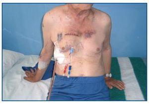
This procedure was carried out successfully in 4 patients, and the following complications were attributable to it: three haemothorax episodes, one of which was massive, for which a thoracotomy was performed on each patient and a chest tube was placed for an average of 5 days; the patient with massive haemothorax required transfusion of 5 units of red blood cells, mediastinostomy, ligation of the bleeding vessels, and thoracotomy with chest tube for 7 days (figures 1 and 2).

Subsequently, the patients were taken for chronic haemodialysis and progressed satisfactorily, without complications attributable to the procedure. Their last average Kt/V was 1.45 and one patient completed 36 months of using the catheter (figure 3).


DISCUSSION

As the population of CKD patients on haemodialysis therapy ages, it becomes increasingly difficult to obtain satisfactory access through which to provide their therapy. A-V fistulas have the tremendous advantage of permitting multiple punctures over a long period of time, but in a group of patients, mainly diabetics, it becomes impossible to place them. The same is true with A-V prosthesis placement. In this group of patients, the use of either temporary or indwelling central venous catheters, inserted through various sites, becomes necessary.

The internal jugular vein approach is the most commonly used due to its easy puncture and the low rate of complications.1 The subclavian route is not recommended, since it results in high stenosis and thrombosis rates, which subsequently prevent the use of the upper extremity for the creation of A-V fistulas.2 The axillary and innominate veins can also be used for the placement of central catheters, but they require medical personnel familiar with those puncture procedures in order to be utilized.3,4 The infradiaphragmatic approach offers several routes: the femoral, with easy access but with the disadvantage of a high thrombosis and infection rates;5 on the other hand, the transhepatic and translumbar routes6,7 are technically more difficult. At the supradiaphragmatic level, two final approaches allow for catheter implantation: the intracardiac and the right parasternal routes, each of which requires the use of general anaesthesia and anterior thoracotomy to access the puncture site. In the intracardiac access the right atrium is punctured, with later placement and tunneling of the indwelling catheter.8

Right parasternal access was first described in the year 2002 by Archundia et al,9 who performed it on a patient who had exhausted all supra- and infradiaphragmatic vascular access points. In this technical report, 3 more patients are being reported, without establishing its long-term outcomes.

The lack of literature indicating the use of this route after its initial introduction is particularly curious.

In our area, about 25% of patients on haemodialysis therapy require the use of indwelling catheters for haemodialysis, which has forced us to employ the vast majority of known routes for both supra- and infradiaphragmatic catheter, including femoral, iliac, and translumbar catheters. Nephrologists perform the aforementioned percutaneous procedures, the majority with ultrasound guidance, while fluoroscopy or computed axial tomography is used only for translumbar placement. When it comes to catheters such as those presented in this case, vascular or thoracic surgeons must be involved. Otherwise, such placement would be impossible, since this medical group possesses the requisite skills and knowledge of intrathoracic anatomy.

Based on our experience, it has been possible to achieve satisfactory placement of 4 catheters in the superior vena cava for 36 months. It is important to note that surgical complications are common, despite the experience of the surgical team with thoracic surgeries. Our recommendation is to always rely on experienced surgeons and to be alert to the occurrence of complications in order to resolve them quickly and avoid fatal consequences.

Table 1. Patient characteristics and outcomes

Figure 1. Patient D on second post-operative day.

Figure 2. PA chest x-ray of patient D.

Figure 3. Patient 36 months after catheter placement.

Bibliography
[1]
Restrepo CA, Chacón JA, Villota DM. Safety related to the implantation of jugular catheters for hemodialysis and usefulness of chest X rays post procedure. Acta Med Colomb 2008;33:68-74.
[2]
Mansfield PF, Hohn DC, Fornage BD, Gregurich MA, Ota DM. Complication and failures of subclavian-vein catheterization. N Engl J Med 1994;331:1735-8. [Pubmed]
[3]
Restrepo Valencia CA. Axillary catheter for hemodialysis, an alternative vascular access. Nefrologia 2008;28:77-81. 4. Restrepo Valencia CA, Buritica Barragán CM. Placement of vascular access catheters for haemodialysis in the innominate vein: a littleused approach. Nefrologia 2009;29:354-7.
[4]
Maya ID, Allon M. Outcome of tunneled femoral hemodialysis catheters: comparison with internal jugular vein catheters. Kidney Int 2005;68:2886-9. [Pubmed]
[5]
Stavropoulos SW, Pan JJ, Clark WI, Soulen MC, Goldberg RDS, Itkin M, et al. Percutaneous transhepatic venous access for hemodialysis. J Vasc Interv Radiol 2003;14:1187-90. [Pubmed]
[6]
Rodríguez-Cruz E, Bonilla M. Percutaneous translumbar inferior vena cava catheter placement for long term hemodialysis treatment. Pediatr Nephrol 2007;22:612-5. [Pubmed]
[7]
Agrawal S, Alaly J R, Misra M. Intracardiac access for hemodialysis: A case series. Hemodialysis Int 2009;13:S18-23. 9. Archundia AG, Mendoza AC, Manrique MN, Figueroa SA. A method to insert a haemodialysis catheter by parasternal access. Nephrol Dial Trasplant 2002;17:134-6.
Download PDF
Idiomas
Nefrología (English Edition)
Article options
Tools
es en

¿Es usted profesional sanitario apto para prescribir o dispensar medicamentos?

Are you a health professional able to prescribe or dispense drugs?