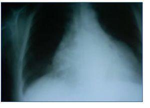
Dear Editor, Microscopic polyangiitis is a necrotising vasculitis primarily affecting the glomeruli (vasculitis limited to the kidney may be the only manifestation), and secondly, the lungs, but it is a systemic process that may affect small blood vessels in any part of the body. High blood pressure is the most common cardiovascular manifestation. We present the case of a patient with sub-acute deterioration of renal function and affected pericardium who responded well to steroid treatment. This 60-year old female patient with a history of diabetes mellitus that was diagnosed three years before, with no microangiopathy or macroangiopathy and high blood pressure during ten years which was treated with carvedilol, hydrochlorothiazide and valsartan. One year before, her creatinine level was 0.8mg/dl, GFR (MDRD) > 60 ml/min/1.73m, and systemic/urine sediment showed no abnormalities. In the six weeks prior to admission, she reported progressive weakness, followed by fever and dry cough. She did not present dyspnoea, chest pain, or diuresis alterations. The examination revealed blood pressure of 190/80mmHg, breathing well, with high jugular vein pressure, rhythmic cardiac sounds with pericardial rub, normal bilateral hemithorax ventilation and no oedema. The analysis showed haemoglobin levels of 7.7mg/dl, urea 217mg/dl, creatinine 4.5mg/dl, potassium 3.6mEq/l, 98% baseline saturation; in the urine; proteinuria of +++ and 60 red blood cells/field. The ECG was normal. The chest radiography showed severe cardiomegaly (figure 1). An emergency echocardiography showed a moderate-to-severe pericardial effusion. Ultrasound showed no kidney alterations. Treatment was started with doses of steroids at 1mg/kg/day, and the patient’s state improved considerably in a few days. With regard to the laboratory results, the direct Coombs test was negative, haptoglobin was 4g/l, LDH 370U/l, C3 and C4 within the normal range, ANA was negative, ANCA was positive, with a p-ANCA pattern. Reactivity analysed by the ELISA test corresponded to antimyeloperoxidase of 146U/ml. Proteinuria was 2g/24 hours. The percutaneous renal biopsy of a total of 17 glomerules was compatible with focal proliferative glomerulonephritis, with crescents at 50% and significant tubulointerstitial involvement. A dose of IV cyclophosphamide was recommended, and the echocardiography was repeated (a day before cyclophosphamide bolus administration) with no signs of the pericardial effusion. After a stay of 18 days, she was discharged with a creatinine value of 1.8mg/dl. In small-vessel vasculitis, it is possible to have primary or secondary cardiac involvement relating to high blood pressure, immunosuppressants, the inflammatory state and the kidney disease that can contribute to developing cardiac hypertrophy, bacterial endocarditis and coronary disease.1,2 As a primary manifestation of the disease, cardiac involvement is more frequent in Churg-Strauss disease, where 13-47%1,3 of all patients have a myocardial, endocardial or pericardial lesion. With regard to the incidence rate for pericardial involvement, whether in the form of acute pericarditis and pericardial effusion or as chronic constrictive pericarditis, its range is estimated between 8 and 32% in Churg-Strauss syndrome and 19% in Wegener’s granulomatosis.5 With regard to microscopic polyangiitis, its unusual presentation as a pericardial effusion caught our attention, and in fact, the literature we consulted contained no references to such an event. Finding it in inflammatory state and the kidney disease that can contribute to developing cardiac hypertrophy, bacterial endocarditis and coronary disease.1,2 As a primary manifestation of the disease, cardiac involvement is more frequent in Churg-Strauss disease, where 13-47%1,3 of all patients have a myocardial, endocardial or pericardial lesion. With regard to the incidence rate for pericardial involvement, whether in the form of acute pericarditis and pericardial effusion or as chronic constrictive pericarditis, its range is estimated between 8 and 32% in Churg-Strauss syndrome and 19% in Wegener’s granulomatosis.5 With regard to microscopic polyangiitis, its unusual presentation as a pericardial effusion caught our attention, and in fact, the literature we consulted contained no references to such an event. Finding it in this case was achieved by hearing the pericardial rub; together with a compatible radiography, this led us to order the echocardiography that confirmed our diagnosis. She began steroid treatment early, with an excellent response. As a result of this experience, we must consider the possibility of pericardial involvement in this type of vasculitis. Proper examination and evaluation of simple complementary tests that are available in every hospital continues to be the fundamental pillar of a correct diagnostic and therapeutic approach.
Figure 1. Cardiomegaly without other signs suggesting heart failure.


