Dear Editor,
Calcific uremic arteriolopathy (calciphylaxis) has an incidence of 1% and prevalence of 4% in the dialysis population. The pathogenesis is not clear and the different treatments (control of Ca-P metabolism, parathyroidectomy, adequate management of wounds, hyperbaric chamber, non-calcium based binders, bisphosphonates, sodium thiosulfate, etc.) did not show any improvements in the prognosis.
The clinical case of a male patient aged 55 diagnosed with Chronic Renal Failure (CRF) secondary to diabetic nephropathy and arterial hypertension is presented. The patient was admitted to Haemodialysis in May 2003.
He had antecedents of diabetes type 2 treated with oral hypoglycaemic drugs and then NPH insulin.
Nonproliferative diabetic retinopathy. Obesity (Body Mass Index [BMI] 36.) Dyslipidaemia. Ex smoker (2 packets a day.) Long-term AHTN (20 years) treated with Angiotensin Converting Enzyme inhibitors (ACE inhibitors) and calcium channel blockers. Peripheral vasculopathy without amputation (Doppler study of the lower extremities with distal compromise and parietal calcifications in bilateral arterial vessels.) No Deep Vein Thrombosis (DVT.) Echocardiography and valvular Doppler with slight deterioration of the RVSD and calcifications in the aortic and mitral valves.
From 2003 to 2005, the patient presented with slight-moderate secondary hyperparathyroidism with hyperphosphataemia that was difficult to treat due to lack of compliance with diet and phosphate binders. A diet low in P was indicated with proteins and calcium binders (first calcium carbonate, then calcium acetate or aluminium sporadically.) Normocalcaemia. KT/V sp greater than 1.4 (15 hrs HD/week) with calcium dialysate 3mEq/l.
Laboratory Data: average haematocrit 35%; haemoglobin 11g/dl; Alb. 3.4g/dl; Ca 8.7mg/dl; P 7.3mg/dl; PTH 538pg/ml; FAlc 350 IU.
The patient was treated, on a discontinuous basis, with oral calcitriol, which was then suspended due to hyperphosphatemia.
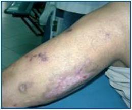
In 2006 the patient presented with symmetric ulcers in the lower distal extremities (legs and feet), pruritic and very painful. Some advanced with eschar and bacterial overinfection due to scatching. The patient was treated with local antibiotic and systemic antibiotics.
Collagen disease tests and coagulation studies were normal, cryoglobulin and anticardiolipin negative.
Skin biopsy showed necrosis of the superficial dermis and localised deposits of calcium in the middle arteriolar layer compatible with calciphylaxis.
Parathyroid hormone (PTH) levels were greater than 800pg/ml and the patient presented with hyperphosphatemia with normocalcaemia. Ultrasound of parathyroids only detected the lower glands. Scintography of the parathyroids with Tc99m and sestamibi detected hypercaptation in three glands.
Patient with caliphylaxis, PTHi greater than 800 and hight CaxP. Daily dialysis was indicated with dialysate calcium 2.5mEq/l. The patient did not improve, due to the persistence of injuries, and subtotal parathyroidectomy was indicated in 2006.
At the end of 2006, the lesions in the lower extremities persisted with PTHi 280pg/ml (range 150-435) and hyper-P. This was interpreted as persistence of HPT.
In February 2007, sevelamer was started, 6 capsules a day, daily dialysis and solution low in calcium (2.5mEq/l), low phosphate diet Oral ibandronate was added, 150mg/month.
Calcaemia controls remained within normal ranges. The lesions in the lower extremities improved and scarred after six months. However, the peripheral vasculopathy progressed and developed into dry gangrene.
Different results with the use of bisphosphonates are described in the literature. They have a potent inhibiting affect on osteoclastic activity and bone resorption, reducing vascular calcification. They also have an inhibiting affect on proinflammatory cytokines, allowing an improvement in the clinical picture.
Figure 1.


