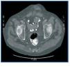
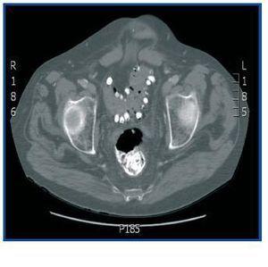
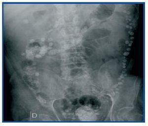
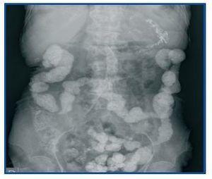
Dear Editor, Lanthanum carbonate is a calcium and aluminium-free phosphorus binder that has recently come to market in Spain. It is a heavy, non-toxic metal that is not absorbed by the intestine. The substance’s package leaflet in our country does not allude to the phenomenon of its appearance in radiology images. This is not the case in the USA’s version, which states that “radio-opaque images may appear in abdominal radiographies of patients who consume lanthanum”.1 The most commonly reported adverse reactions were gastrointestinal, but the clinical trials did not include patients with intestinal obstructions or inflammatory intestinal disease.2 We present the case of a 58-year old man with pan-colonic diverticulosis and frequent diverticulitis episodes with CKD secondary to diabetic nephropathy who began a periodic haemodialysis programme in April 2001. He was admitted in July 2008 for fever and abdominal pain. An emergency abdominal CT ruled out signs of diverticulitis, but the radiologist reported “remnants of contrast in the entire colon and terminal ileum” (figure 1), which was confirmed by a simple abdominal x-ray (figure 2). Our patient had not received any radiological contrast at any time, but he had been receiving treatment with 3000mg lanthanum carbonate daily for severe hyperphosphataemia since February of that year, with excellent lab results and good clinical tolerance up to that moment. The final diagnosis was sepsis due to Enterococo avium, most likely of intestinal origin. Since there were no other findings in the imaging tests that could explain the abdominal pain, lanthanum treatment was discontinued, after which the patient remained asymptomatic. With a view to studying the findings, a simple abdominal radiograph was taken in another patient receiving the same dose of that metal and who had not had any digestive symptoms. The deposit was also observed throughout the contour of the colon, but showed a different radiological pattern (figure 3). References in the literature describing this phenomenon are scarce and contain various explanations. According to our research, the first radiological image attributed to lanthanum consumption was shown by Cerny and Kunzendorf3 in 2006. In this case, the drug was discontinued because after seeing the radiography, doctors felt that the patient’s abdominal pain could be related with the lanthanum. Other cases were subsequently reported.4 David et al.5 interpreted the radiograph as an intestinal deposit of calcium phosphate stones that prove lanthanum’s effectiveness as a binder, and even suggest that such an image could be used as a test of therapeutic compliance. That theory is refuted by Pafcugova et al.,6 who showed that the tablets themselves inside a vial are radio-opaque, in absence of calcium or phosphorus. However, given our limited experience with the use of this drug, particularly in Spain, it is still not clear what the radiographical distribution pattern is in the abdomen, or whether it can be observed in all patients receiving this medication. Vrigneaud et al.7 studied 13 patients treated with lanthanum. In six, the abdominal radiograph is completely normal, while in the rest, the radioopaque deposit may be observed. However, in contrast with the habitual images with opaque areas smaller than one centimetre distributed regularly throughout the contour of the colon, we found two cases in which the areas were larger and irregularly distributed along the digestive tract. These were in one patient who did not chew the tablets correctly, and another patient with colonic diverticulosis whose profile suggests the material was deposited in the diverticuli, as with the present case. Our conclusion is that although little data exists about the radiological behaviour of intestinal lanthanum carbonate deposits, it is necessary to know that it is radio-opaque. Furthermore, it is likely that the drug should be used cautiously in patients with intestinal diseases. In addition, we do not know its elimination time, which is crucial information when it comes to performing other imaging studies without interferences. And lastly, we believe that all of the above should be included in the drug’s technical leaflet printed in Spain.
Figure 1. Abdominal CT without contrast showing the lanthanum carbonate deposit in the sigmoid colon diverticuli and rectum.
Figure 2. Simple abdominal radiography without contrast showing multiple onecentimetre sized opaque points irregularly distributed along the contour of the colon, which are lanthanum deposits in the diverticuli.
Figure 3. Simple abdominal radiography without contrast showing radio-opaque material distributed along the entire contour of the colon. This image is from another patient receiving the same dose of lanthanum, but with no abdominal symptoms.


