Dear Editor,
Patients with acute renal failure due to pauci-immune necrotizing and crescentic glomerulonephritis with antinuclear antibody (ANCA) seropositivity can present with positive lupus serologies.1 On the other hand, patients with lupus nephritis present with ANCA seroconversion in 20% of cases. The fact that systemic lupus erythematosus (SLE) and positive myeloperoxidase (MPO) ANCA titers with kidney involvement can present with scant subendothelial deposits in the kidney biopsy, may suggest a forme fruste of lupus nephritis or a concomitant renal vasculitis with neutrophil priming.
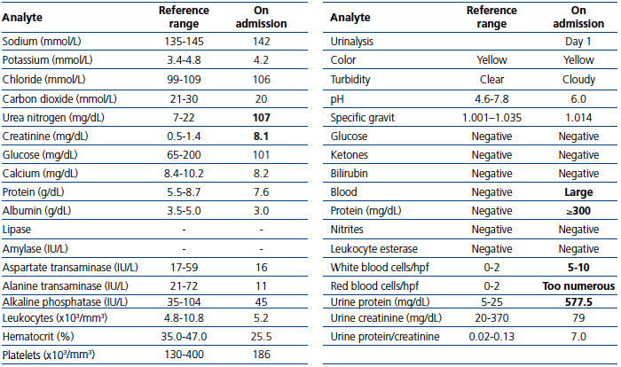
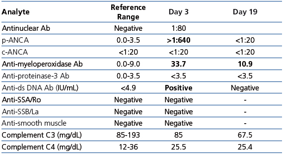
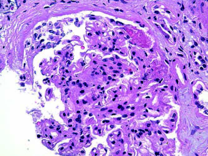
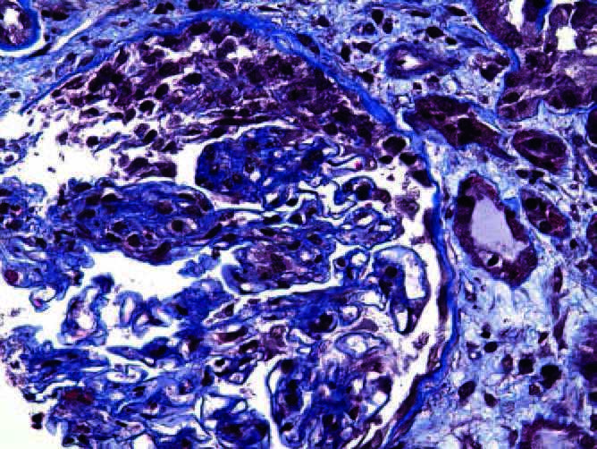
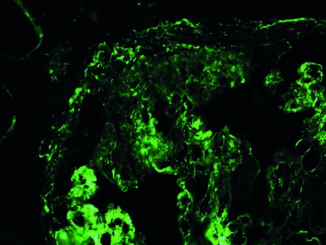
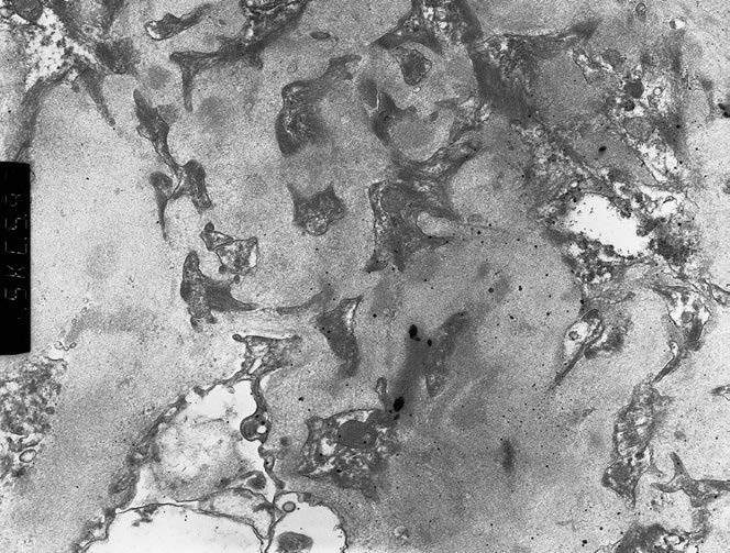
A 77-year-old man with chronic kidney disease due to hypertension, presented with hematuria, nausea, and vomiting and red discoloration of urine. Laboratory data Table 1, serology tests Table 2. Renal ultrasonography unremarkable. Patient developed hemoptysis. Chest radiograph revealed bilateral diffuse airspace opacities. Intravenous methylprednisolone was administered. The patient received hemodialysis. Renal biopsy showed mesangial hypercellularity (Figure 1), crescents (Figure 2), segmental necrosis (Figure 1). There was moderate tubular atrophy an occasional eosinophil. Immunofluorescence microscopy demonstrated granular IgG (1+), C3 (2+), and C1q (1+) deposition in the mesangial areas and glomerular basement membranes (Figure 3). EM showed numerous electron-dense deposits in the mesangial areas and few subepithelial and subendothelial electron-dense deposits (Figure 4). Focal effacement of podocyte foot processes was noted. Histological diagnosis: immune complex-mediated necrotizing and crescentic glomerulonephritis.
Patient received pulse Rituximab and cyclophosphamide. The hospital course was complicated by hypoxic respiratory failure. Folow up computed tomography of the chest showed a right lower lobe pulmonary embolism. Anticoagulation with heparin was initiated. Serological tests were repeated (Table 2) and showed normalization of p-ANCA (<1:20) and anti-double-stranded DNA antibodies. Anti-MPO antibodies were reduced at 9.8U/mL after induction therapy. The patient expired.
Pauci-immune necrotizing and crescentic glomerulonephritis (GN) due to the activation of neutrophils by ANCA, differs from lupus nephritis in that glomerular necrosis and crescent formation occurs in the absence of cellular proliferation and in the presence of scant immune-complex deposition. ANCA are implicated in the pathogenesis targeting cytokine-primed leukocytes that expressed MPO or proteinase 3 (PR 3) instead the white blood cell surface.2
Lupus nephritis is an immune complex-mediated renal disease were the formation of glomerular immune deposits results in complement activation, leukocyte infiltration, cytokine release, cellular proliferation, crescent formation, and necrosis under certain circumstances. The final result is glomerular scarring.1 There are cases of lupus nephritis in which focal or diffuse glomerular necrosis and crescents occur without substantial subendothelial deposits.3 Patients with lupus nephritis IV-S (2003 International Society of Nephrology/Renal Pathology Society classification/(endocapillary or extracapillary GN involving >50% of glomeruli with segmental lesions) had extensive fibrinoid necrosis and less immune-complex deposition findings resembling a pauci-immune GN at times.4 Approximately 20% of patients with SLE have ANCA positivity by immunofluorescence microscopy (IF), mainly with a perinuclear (p-ANCA) pattern.5 Antinuclear antibody seropositivity by enzyme-linked immune-sorbent assay (ELISA) is less frequent, and target antigens are most commonly lactoferrin (LF), cathepsin G, and MPO.5 Galeazzi et al. evaluated 566 patients with SLE and found ANCA positivity by immunofluorescence microscopy in 16.4% of them including 15.4% p-ANCA and 1% c-ANCA pattern. By ELISA, 9.3% had MPO-ANCA positivity and 1.7% had PR3-ANCA positivity.6 There is difficulty in distinguishing p-ANCA from ANA by immunofluorescence microscopy.7 There are also conflicting reports on biological significance of ANCA in patients with SLE. Antinuclear antibody positivity has been associated with the presence of nephritis, particularly diffuse proliferative lupus nephritis, as well as anti-dsDNA antibodies.8 While other reports have failed to show a correlation between ANCA and organ involvement.9 Nasr et al. evaluated a cohort of ten patients with SLE, ANCA positivity and renal biopsy findings of lupus nephritis and ANCA-associated GN. All biopsies exhibited necrosis and crescents with no or rare subendothelial deposits.7 Nine patients had p-ANCA positivity by IF. The high incidence of MPO-ANCA seropositivity in patients with SLE, raises the possibility that the findings are not coincidental occurrence of two unrelated diseases. One condition might trigger the other one or vice versa. Systemic lupus erythematous may facilitate MPO autoantibody formation by promoting neutrophil degranulation and priming neutrophils to increase surface expression of MPO.7 On the other hand, the association of autoantibodies to MPO in drug-induced SLE has been reported independently.10 There are conflicting reports on the correlation between the presence of ANCA in SLE and clinical features. Some reports show no correlation between organ involvement and the presence of ANCA, whilst others report a link. In the largest population of patients studied, Galeazzi et al. reported significant positive correlations between IF ANCA and venous thrombosis as well as serositis.6 In this case, our patient presented with an episode of pulmonary embolism, which is consistent this hypothesis. Here, we presented a patient with systemic vasculitis with rapid progressive glomerulonephritis and necrotizing and crescentic changes. Lupus serologies probably represented an autoimmune response to the antinuclear antibody activity. The fact that he had few sub endothelial deposits and lack of hypocomplementemia goes against activation of immune complexes due to SLE. In the other hand, the possibility of a simultaneous ANCA/lupus nephritis involvement represents an interesting hypothesis.
Conflict of interest
The authors declare that they have no conflicts of interest related to the contents of this article.
Table 1. Laboratory data
Table 2. Serologic tests
Figure 1. H&E: mesangial hyeprcellularity and necrotizing crescentic glomerulonephritis
Figure 2. Tricrome: shows cellular crescent (stained red) extending from 11 to 2 o'clock postion
Figure 3. IF for C3: granular predominantly mesangial deposits
Figure 4. EM: electron dense immune complex deposits in glomerular mesangium (high power)


