Dear Editor:
Renal transplant (RT) patients have a higher incidence of thrombotic events and an increased risk of recurrence after the withdrawal of anticoagulation. Thrombosis of the allograft vein is a well-described complication of renal transplantation. It can occur early after transplant, related to surgical technical complications or many years post-transplant associated to multiple inciting factors. The treatment includes surgery, thrombolytics and anticoagulation.
We present two cases of late allograft venous thrombosis with different treatments and outcome: conventional hipocoagulation leaded to renal failure but surgical thrombectomy allowed patient improvement and renal function recovery. Based on the cases, a review of the literature about pathophysiology, clinical presentation, diagnosis and treatment options of late venous thrombosis of renal allograft was made.
RT patients have a higher incidence (ranging 0.6-25%) of thrombotic events1,2. Thrombosis of the allograft vein is a well-described early complication3, usually associated with acute rejection or surgical complications4. The typical presentation is that of a sudden painful and swollen allograft, haematuria and oliguria with deterioration of graft function4,5. Partial vein thrombosis presents as a late event, with chronic oedema and progressive deterioration of renal function6.
Diagnosis can be made by Doppler ultrasound, computed tomography (CT) or magnetic resonance venogram7 and the treatment includes surgery, thrombolytics and anticoagulants7.
The authors present two cases of late allograft venous thrombosis with different treatments and outcome.
CLINICAL CASES
Case 1
A 63-year-old man, with chronic renal failure (CRF) secondary to adult polycystic kidney disease (APKD), was submitted to RT in 1988 and treated with cyclosporine (CsA), azathioprine (AZA) and prednisolone (P). Nineteen years after RT, serum creatinine (Cr) increased to 2.5 mg/dl and nephrotic proteinuria was documented. In 2007, chronic allograft nephropathy (CAN) was confirmed. One year latter, a rectal adenoma was diagnosed and after four months (on March 2009), he had acute diverticulitis complicated by peritonitis and needed surgery.
On July 2009, he was admitted with painful oedema of the right leg with one week of evolution. Doppler revealed femoral vein thrombosis and partial thrombosis of allograft vein, iliac and inferior vena cava (IVC). Renal function had declined (Cr: 5.84 mg/dl) and serum albumin was reduced (2.68 g/dL). Pulmonary embolism was excluded and anticoagulation with low molecular weight heparin (LMWH) was started, followed by accenocumarol. Renal function deteriorated and one week latter he started haemodialysis. The study for other neoplasms was negative. Three months latter, he is asymptomatic but remains on haemodialysis.
Case 2
A 58-year-old man, with CRF secondary to APKD, was submitted to RT in 1993. He was treated with CsA, AZA and P and renal function stabilized on Cr: 1.8 mg/dl, without proteinuria. Posttransplant erytrocytosis was documented in 1996 and treated with phlebotomies.
On May 2009, he was admitted with thrombosis of right popliteal vein. He had erytrocytosis (Hb: 18.3g/dL) and deterioration of renal function (Cr: 2.2 mg/dl). Anticoagulant treatment was maintained for 6 weeks, with improvement.
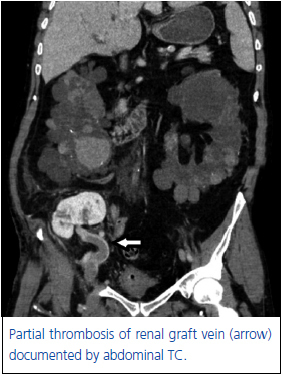
Three months latter, he was readmitted with oedema of right leg with two days of evolution. He maintained erytrocytosis (Hb: 16.9 g/dL) and allograft dysfunction (Cr: 2.02 mg/dl). Imagiological studies revealed thrombosis of femoral vein with extension to allograft and iliac veins, without involvement of IVC (figure 1). No neoplasic disease was found.
He was treated with heparin without improvement, and started haemodialysis on the 3rd day. Surgical thrombectomy was preformed and, one week latter, renal function recovered (to Cr: 1.8 mg/dl). He was discharged under oral anticoagulation and two months latter, he is asymptomatic with stable renal function (Cr: 1.79 mg/dl).
DISCUSSION
Early allograft venous thrombosis accounts for one third of all graft losses within the first three postttransplant months5. Thrombosis occuring several months after RT is rare8 and is associated to inciting factors5,7. RT patients have persistent hypercoagulable state that may play a role in latter thrombotic events (TE)2. Clotting activation is multifactorial, with classic risk factors associated to specific ones related to RT1-4.
Allograft vein thrombosis is more frequent with some therapies, particularly with OKT3 and high doses of steroids4,9. CsA role remains controversial5,9. Our patients were treated with low doses of immunosuppression and is unlikely that therapy alone caused thrombosis.
Recurrent or de novo glomerulonephritis with proteinuria superior to 2 g/day10 (even without nephrotic syndrome) generates hypercoagulable states1,4,7 and neoplasms increase the risk of thromboembolism by nearly five times in RT patients7.
Late renal vein thrombosis (RVT) was described following surgery, related to immobilization or hypovolemia, and associated with compression of the allograft vein4,8. The first patient had nephrotic proteinuria, a neoplasic lesion and was recovering from surgery with prolonged immobilization. Either polycystic kidneys or adhesions could compress allograft vein and act as predisposing factors.
Posttransplant erythrocytosis affects 10-15% of RT recipients11 and was pointed as the inciting factor for thrombosis2,5 in the second patient.
Few weeks after the first episode, we confirmed recurrence of venous thrombosis with extension to allograft vein. After anticoagulants withdrawal, the risk of TE recurrence is near 48% in RT recipients3, which is 10 times higher than in normal population2,3.
The treatment of RVT includes anticoagulants, thrombolytics and thrombectomy. Most cases of early post-surgical RVT are treated with thrombectomy, but in the late posttransplant it has low success rate12. Some authors advice surgical thrombectomy only when a surgical cause is identified and there aren’t adhesions that make surgery unsafe7. The first 10-14 posttransplant days are considered the timing for an open approach. Beyond that time, a percutaneous approach is recommended7.
Mechanical thrombectomy can lead to pulmonary embolism (PE)13, specially if the thrombus has extension to the IVC, as in our case 1.
Alternative treatments for late RVT include anticoagulation with heparin/LMWH or drug-induced thrombolysis7. Thrombolytic agents have proved better results, with complete lysis in 40-60% of patients, compared to 10% of those treated with heparin14.
Thrombolytics are more efficient when thrombi are less than 5 days-old15, but the most effective agent and the optimal duration of treatment remain uncertain7.
A combined approach of percutaneous mechanical and chemical thrombectomy has been used7,13. It is advocated in RVT beyond the second week posttransplantation or when prolonged thrombolysis is contraindicated7,13.
In our first case, post-peritonitis adhesions made surgical approach difficult, the organized thrombus reduced thrombolysis efficacy and the high probability of irreversible damage (in a graft with CAN) contributed to the decision for a conservative treatment. In our second patient, thrombectomy was really efficient, allowing allograft recovery.
In conclusion, renal vein thrombosis in late pos-transplant period is not an indication to graftectomy neither a definitive evidence of graft failure. Therapies such as thrombolysis or thrombectomy must be considered, as they may allow better outcomes.
Figure 1. Thrombosis of renal graft vein.


