To the Editor,
Hydrothorax is a peritoneal dialysis complication, associated with an increase in intra-abdominal pressure,1 which on very rare occasions has been reported to be related with peritonitis episodes.2,3 We present the case of a patient who, after having had other complications related with an increase in intra-abdominal pressure, developed hydrothorax during the course of a peritonitis episode.4
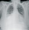
She is a 76-year-old woman, with stage 5 chronic kidney disease with interstitial nephropathy. Over the past three years, she has been receiving continuous ambulatory peritoneal dialysis, with three daily 2,000ml refills. Over this period, she has not suffered peritonitis nor has she undergone surgery. In a routine check-up, a small non-incarcerated umbilical hernia and an abdominal hernia were detected, the latter measuring approximately 6cm in diameter in the left paramedian line and confirmed by CT scan and ultrasound. The peritoneal dialysis was not affected by these anatomical alterations, so after surgical evaluation a conservative stance was maintained, modifying the dialysis rate to four daily 1,500ml refills. She returned to the hospital six months later with cloudy fluid, fever and mild abdominal pain, with a 5 hour evolution. In the days before, she had suffered mild diarrhoea. The peritoneal effluent cell count was 4,570cells/ml (98% PMN). Diagnosed with bacterial peritonitis in peritoneal dialysis, she was admitted and put on intraperitoneal antibiotic therapy, following our centre’s protocol: vancomycin, ampicillin and tobramycin. On the following day, the fluid was still cloudy and cell count was 9,850cells/ml (94% PMN); the peritoneal dialysis balance showed a gain of 1,200ml and the abdominal pain persisted. Thirty-six hours after admittance, she suffered sudden dyspnoea. The patient presented a generally poor state: she was pale, sweaty, tachypneic and had difficulty breathing. Oxygen saturation was 90%. The respiratory auscultation displayed a reduced breath sounds in the right hemithorax. The chest X-ray displays a moderate right pleural effusion (Figure 1). A short hypertonic refill is administered with no improvement. Due to paroxysmal breathing difficulty and the X-ray results, thoracocentesis was performed, which drained 2,000ml. The pleural fluid was a glucose concentration of 269mg/dl, much higher than the plasmatic concentration (110mg/dl), which confirmed hydrothorax. Improvement was significant after the pleural tap. The peritoneal dialysis was suspended and on receiving the peritoneal fluid culture, positive for Bacteroidesfragilis, intravenous antibiotic therapy was started with meropenem and metronidazole. The abdominal CT scan ruled out perforation and other intra-abdominal pathologies. After two weeks of treatment, she was discharged and is currently on regular haemodialysis.
Hydrothorax consists of peritoneal fluid passing to the pleural space through a peritoneal-pleural structural defect, and can be either congenital or acquired.1-3,5-8 Its prevalence is hard to estimate and varies from 1.6% and 2.9% to 10%, according to studies.3,5,6 It appears more commonly in females and in the right hemithorax,5 with a tendency to manifest at the start of the peritoneal dialysis technique, although it is not unusual for it to appear months or even years later. There are two factors that are combined in its aetiology and pathogenesis: on the one hand, the increase in intra-abdominal pressure and, on the other, the existence of continuity solutions in the pleuro-peritoneum, either congenital or acquired.7,8 Peritonitis episodes can also worsen congenital diaphragmatic defects, although we have only found two reported cases and both are quite old, confirming the scarceness of this possibility.2,3 Clinical results are diverse, depending on the amount of pleural effusion. In some studies, up to 25% of the cases go unnoticed.5 In others, there may be paroxysmal dyspnoea, thoracic pain and coughing, together with a decrease in ultrafiltration. The examination showed abolition of breath sounds, dullness on percussion and no transmission of vocal vibrations. Diagnosis was via chest X-ray and pleural fluid analysis (with higher glucose concentration than plasmatic). Stainings can be used (methylene blue) and peritoneography with contrast medium to reveal the existence of pleuro-peritoneal communications, although these methods can cause peritoneal irritation. Currently, the most common technique applied is peritoneal scintigraphy with technetium-99.4,7 Several therapeutic options have been reported: transitory haemodialysis with a return to peritoneal dialysis with low volumes or with a cycler, pleural tap, pleurodesis with chemical substances or autologous blood transfusion and direct surgical repair or thoracoscopy.3,5-8 Nonetheless, treatment results are not very encouraging and in a high proportion of cases, the patient is definitively transferred to dialysis.3,5-7
In our patient, advanced in age, the increase in intra-abdominal pressure due to the peritoneal dialysis gave rise to, firstly, a hernia. This increase in pressure with the structural weakness of the diaphragmatic parietal peritoneum caused by peritonitis generated pleuro-peritoneal communication, giving rise to hydrothorax. Given these complications haemodialysis was chosen as the most appropriate extrarenal purification technique, although it was against the patient’s will.
Figure 1. Hydrothorax


