Infusion/drainage problems are defined as a slowed flow, hampered or prevented by causes related to the catheter itself and not to the functioning of the peritoneum as a dialysis membrane. The occurrence of these problems ranges between 5% and 20% and is often associated with the technique used fro the placement of the catheter. They are more common in cases of implantation by laparoscopy.1
Let us look at the case of a patient suffering catheter flow problems who was diagnosed with partial catheter obstruction by fluoroscopic peritoneography and whose transfer to haemodialysis was avoided.
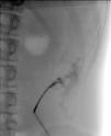
The patient was a 17-year-old female with CKD secondary to IgA nephropathy, on an automated peritoneal dialysis (APD) program since April 2015 through a double-cuff silicon catheter and with a pigtail end implanted by open surgery. She had experienced no mechanical or infectious problems related to the technique, and maintained a residual diuresis of 1500ml per day. She came to the unit complaining that she could not perform dialysis due to multiple concerns. She did not report constipation, although she had only one small bowel movement a day. An exchange was performed at the unit, in which infusion was very slow and drainage heavily obstructed. A push and suck maneuver was applied, checking the permeability of the catheter. The most common causes of infusion problems are catheter buckling and blockage of the catheter lumen/hole due to fibrin. And the causes of drainage problems are either of the two previously mentioned, plus poor catheter placement, obstruction due to omentum entrapment and constipation.2 An abdominal X-ray was performed showing the distal end of the catheter displaced in the left iliac fossa and abundant gases and stool remains. For diagnosis, the simple X-ray identifies the position of the catheter in the abdominal cavity. Enemas and laxatives were prescribed and walking was recommended. Patient showed no volume problems or uremic symptoms. 48h later the patient returned without any resolution of the problem. The abdominal X-ray was repeated which showed no changes in terms of the positioning of the catheter, and so the alpha maneuver was performed. Poorly placed catheters can be repositioned using a vascular guide wire (alpha maneuver).3 This can resolve some 50–80% of cases, although only 33% will achieve a permanent solution. The catheter was able to be moved down by around 3cm, but the drainage problem persisted. Iodinated contrast medium (10ml) was infused by catheter and fluoroscopy showed that it only came out through the proximal orifices of the catheter (Fig. 1).
In cases where diagnosis is difficult, performing a CT or MR peritoneography, or the less-used fluoroscopy, can diagnose virtually all of such types of complications, ruling out leakage.4,5
As for treatment, this depends on the case; constipation can be treated with a fiber-rich diet, laxatives or enemas. Nearly 50% of drainage difficulties are resolved using these methods. Where fibrin plugs or strands appear in the effluent, adding 200–500U/l of heparin to the dialysis fluid is beneficial. Where the fibrin causes occlusion of the catheter lumen, as in the case in point, the instillation of 5000U of urokinase may be adopted, which should be maintained for 1h.1 The catheter was sealed with urokinase, achieving acceptable infusion and drainage flows. Since this measure was effective, heparin was added to the subsequent changes at the dose mentioned, the prescription was changed to manual with CAPD and a regular use of laxatives was recommended. The patient was subsequently monitored as an outpatient by telephone. Since the flows improved over time, the patient resumed APD.
Most complications that cause catheter infusion and drainage problems which cannot be resolved with conservative methods can be approached using laparoscopy techniques: repositioning of poorly placed catheters and suture fixation, fibrin occlusion cleaning, clearing obstructions caused by omentum entrapment and omentectomy, or replacement with a self-locating catheter.6
Prevention of these types of conditions requires proper catheter placement and avoiding causes of constipation.
Please cite this article as: Sastre A, González-Arregoces J, Romainoik I, Mariño S, Lucas C, Monfá E, et al. Diagnóstico de obstrucción de catéter peritoneal mediante peritoneografía fluoroscópica. Nefrologia. 2017;37:101–103.


