Journal Information
Vol. 36. Issue. 6.November - December 2016
Pages 583-722
Visits
9668
Vol. 36. Issue. 6.November - December 2016
Pages 583-722
Review
Open Access
Detection of cardiovascular calcifications: Is it a useful tool for nephrologists?
Detección de las calcificaciones cardiovasculares: ¿una herramienta útil para el nefrólogo?
Visits
9668
Jordi Bovera,
Corresponding author
jbover@fundacio-puigvert.es

Corresponding author.
, José Luis Górrizb, Pablo Ureña-Torresc, María Jesús Lloreta, César Ruiz-Garcíaa, Iara daSilvaa, Pamela Changa, Mariano Rodríguezd, José Ballarína
a Servicio de Nefrología, Fundació Puigvert, IIB Sant Pau, RedinRen, Barcelona, Spain
b Servicio de Nefrología, Hospital Universitario Doctor Peset, Valencia, Spain
c Departamento de Nefrología y Diálisis, Clinique du Landy, Departamento de Fisiología Renal, Hospital Necker, Universidad de París Descartes, París, France
d Servicio de Nefrología, Hospital de Universitario Reina Sofía, IMIBIC, Universidad de Córdoba, Córdoba, Spain
This item has received

Under a Creative Commons license
Article information
Abstract
Full Text
Bibliography
Download PDF
Statistics
Figures (1)
Abstract

Chronic kidney disease (CKD) has been used as a model and source of knowledge concerning the mechanisms, clinical relevance and accelerated progression of cardiovascular (CV) calcification, as well as its consequences in clinical practice. However, we know that it is a late secondary ossification phenomenon and only circumstantial evidence is available. In this comprehensive review, we firstly describe the types of CV calcification which affect CKD patients, and we analyse how its presence is directly associated with CV events and increased mortality in these patients. We also justify the use of CV calcification assessment in regular nephrology clinical practice, because CV calcification is an important predictor of clinical outcome in these patients. Consequently, we believe that CV calcification assessment is a tool that could and should be used by nephrologists when making a decision concerning individual patients, consistent with the current trend of an ever-more-personalised therapeutic approach.

Keywords:
Chronic kidney disease
Vascular calcification
Adragao
Kauppila
Agatston
CKD-MBD
Mineral metabolism
Calcium
Phosphorus
Hyperparathyroidism
Resumen

La enfermedad renal crónica (ERC) ha servido de modelo y fuente de conocimiento sobre los mecanismos, la relevancia clínica y progresión acelerada de los procesos de la calcificación cardiovascular (CV), así como de sus repercusiones en la práctica clínica, aunque se trate de un fenómeno tardío y secundario de osificación sobre el que solo disponemos de evidencias circunstanciales. En esta amplia revisión se describen primero los tipos de calcificación CV que afectan al paciente con ERC y se analiza cómo su presencia está directamente asociada a eventos CV y a un aumento de la mortalidad de estos pacientes. Asimismo, justificamos la valoración de la calcificación CV en la práctica clínica nefrológica habitual, al entender que es un predictor importante de la evolución clínica de estos pacientes, y consideramos que la valoración de las calcificaciones CV es una herramienta que puede y debe ser utilizada por el nefrólogo para la toma individualizada de decisiones terapéuticas en un momento en que se requiere cada vez más de una medicina personalizada.

Palabras clave:
Enfermedad renal crónica
Calcificación vascular
Adragao
Kauppila
Agatston
CKD-MBD
Metabolismo mineral
Calcio
Fósforo
Hiperparatiroidismo
Full Text
Introduction

It is currently accepted that chronic kidney disease (CKD) is an independent cardiovascular (CV) risk factor and that mortality rates increase exponentially as kidney function progressively deteriorates.1 Although the first associations between CKD and CV disease date back for more than 40 years, the true scope of the problem only became clear a little over than a decade ago. In this direction several initiatives, among others, by the Spanish Society of Nephrology, have been influential, such as implementation of a systematic estimation of the glomerular filtration rate2–7 enabling early detection of CKD and uncover hidden CKD in Spain.8 Moreover, other recent initiatives have made physicians from other specialties to be aware of the need to diagnose and stage the disease, and provide special care for kidney disease patients; to such extent that consensus documents involving up to 10 national societies have been created in Spain.6,9–11 The growing importance of CKD as a health issue, and especially for the speciality of Nephrology, is illustrated by the important value given to assessment of kidney function in clinical guidelines and publications from other specialities.12–16

In the context of the cardiorenal syndrome17 and with the close correlation between CKD and CV disease, the clinical relevance of CV calcification, even beyond kidney disease patients, is currently being widely debated.13,18–22 CKD has served as a model and source of knowledge about the mechanisms and clinical relevance of the presence and accelerated progression of the arteriosclerosis and vascular calcification processes and their repercussions in daily clinical practice.18,23–27 Knowledge about this condition is advancing at such a pace that it is worth pausing and refreshing this topic, connecting the most basic aspects with the clinical aspects and trying to be objective and realistic when reaching conclusions on the possible strategies for our patients’ care. Therefore, in the first part of this review we will briefly describe the types of CV calcification, associations with both high- and low-bone turnover diseases,24,28 and we will analyse how its presence is directly linked to CV events and increased mortality. The second part will discuss how CV calcification, despite of being a late and secondary phenomenon with only circumstantial evidence available,19,20 is a modifiable risk factor to which we can unfortunately contribute with unwanted iatrogenia.18,29

CKD, cardiovascular risk, and “CKD-MBD”

In addition to the traditional CV risk factors (advanced age, obesity, tobacco use, diabetes, hypertension, dyslipidaemia), a set of non-traditional factors may explain the disproportionate mortality observed in the CKD population. These include a series of modifiable parameters of bone-mineral metabolism disorders such as changes in phosphorus (P), calcium (Ca), parathyroid hormone (PTH), vitamin D, or the fibroblast growth factor 23 (FGF23)/klotho axis, among others30–32; other factor directly or indirectly related include: inflammation, oxidative stress, or changes in the Wnt/β-catenin signalling pathway.19,33–36 The new widely accepted term CKD-MBD (acronym for chronic kidney disease-mineral and bone disorder, with inconsistent Spanish translation39,40) describes the systemic consequences and the organ damage beyond bone caused by altered mineral metabolism in CKD patients.37,38 The CKD-MBD term includes vascular, valve, and extraskeletal calcifications in addition to the biochemical and bone abnormalities. These vascular calcifications are also recognised from a pathophysiological perspective within the new concept of “bone-vascular axis”, which directly relates bone with the CV system. Now we are aware that the bone is an endocrine organ that secretes hormones such as FGF23, sclerostin, or osteocalcin, among others and that is essential part of CKD.41–47 This association affects not only CKD patients, but patients from other specialities.48,49

Anatomic location and histological types of cardiovascular calcification

The presentation of CV calcification may be very heterogeneous. The following types have been described: 1) classical atherosclerosis-atheromatosis, in the context of degenerative changes of the aorta and large elastic arteries, and directly linked to inflammation and dyslipidaemia19,50; 2) medial calcification or Mönckeberg's disease, in the context of concentric thickening of the muscular layer media of the arteries50; 3) cardiac valve calcification; and 4) calciphylaxis or calcific uraemic arteriolopathy.24,50–52 Recently, it has been described a form of vascular calcification limited to the internal elastic lamina,53,54 and some authors have revelled the potential importance of myocardial calcification in inducing electrical disorders/sudden death.55,56

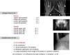
All arteries, even the smallest arterioles, may be affected by the calcification process; veins are rarely involved.50,57–60 For instance, the radial, cubital, and interdigital arteries are regulate the blood flow by changing the tone thanks to a dense layer of vascular smooth muscle cells which can be calcified.27,44,58,59 The wall of iliac and femoral arteries (predominantly but not exclusively made with vascular smooth muscle cells) are also more susceptible to develop calcification than the average.59 All these arteries are assessed by plain x-ray and they are used to calculate the Adragao score58 (Fig. 1). Another method for assessing calcification is the Kauppila score (Fig. 1) that evaluates the abdominal aorta, an elastic artery more susceptible for intimal calcification, given that elastic arteries (such as the subclavian and carotid arteries) have a media layer that contains more elastic fibres than muscle cells.27,44,50,54

Fig. 1.

Adragao Score: Plain x-ray of the hands and pelvis. Determined by the sum of the absence of calcification (0 points), unilateral (1 point) or bilateral (2 points) presence of linear calcifications in each section. It analyses calcification of the iliac, femoral, radial, and digital arteries. The final value ranges between 0 and 8 points (0–4 in the pelvis and 0–4 in the hands).58

Kauppila Score: A lateral abdominal x-ray is performed that includes from the T-10 vertebra to the first 2 sacral vertebra. The aorta is identified as a tubular structure in front of the vertebral column. Only the segments of the abdominal aorta that are in front of the first 4 lumbar vertebrae (L1-L4) are analysed. The score is assigned from 1 to 3 [1: small calcification (1/3 the vertebra length), 2: moderate (2/3), 3: large (affects more than 2/3 of the vertebrae length)] according to the length of each detected plaque. Both the anterior and posterior part of the aorta are taken into account, relating them with the location according to where they are located in front of the L1, L2, L3, or L4 vertebrae. With this scoring, a final score is reached between 0 and 24 points.135

(0.25MB).

Coronary artery calcification (CAC) has been described in 75% of the necropsies in the general population61 and in more than 95% of dialysis patients,62 other prevalence reports are variable (47–98%).63,64 The reported prevalence depends on the population, the vascular region being evaluated, and the sensitivity of the different diagnostic techniques used.63,65,66 Górriz et al. recently described that in CKD patients not on dialysis, besides the coronary calcifications,67 the vascular calcifications detected by hand and pelvic plain x-rays are a solid predictor of hospital-free survival, CV mortality, and overall mortality, even better that aortic calcification.59 This observation has important clinical implications that we will discuss in the second part of this review.29

Vascular and valve calcifications are more common and progress faster in chronic kidney disease: pathogenesis and implications

CV calcification is not a new phenomenon nor it is exclusive to CKD.24,68 It is also common in diabetic patients and is closely correlated with age/ageing.13,27,48 Calcification is not a primary aetiological factor of arterial disease,19 since intimal calcification is part of the natural history of atherosclerosis at its late stages (stage vii), although it is associated with ischaemic CV events. Medial calcification, as an expression of arteriosclerosis, induces artery stiffness and increases the pulse wave velocity which contributes to development of left ventricle hypertrophy, fibrosis, ventricle dysfunction, decreased coronary irrigation during the diastole, and heart failure. The degree of calcification of each lesion has a variable relation with the severity of the associated stenosis; and, the relationship between the degree of calcification in an individual lesion and the probability of the plaque rupturing is unknown. Nevertheless, the presence of any type of vascular calcification is clearly associated with the degree of atheromatosis (which is in turn influenced by multiple factors such as age, dyslipidaemia, diabetes, CKD stage, time on dialysis, etc.), and with CV events, hospitalisation and mortality.27,59,69

The opinion of experts is not uniform,19,70–75 but it has not been confirmed that intimal calcification per se is a risk factor for plaque rupture and, it is well known that inflammation precedes the process of calcification and that they rarely overlap.76,77 However, this calcification seems to be directly associated with CV events.78,79 Even though calcification is a very late secondary phenomenon (progression of initial inflammation and atherosclerosis), there is not convincing evidence that calcification contributes to plaque stabilisation.80,81 Recent literature reveals that local tissue stress could increase due to the presence of juxtaluminal calcification and encrusted calcifications in the fibrous cap.81,82 Similarly, in CKD patients the atheroma plaque composition is characterised by an increase in calcification and a reduction in the amount of collagen which could lead to instability and plaque rupture.83,84 In any case, at least in haemodialysis patients, it must be taken into account that the most common cause of mortality does not seem to be plaque rupture, but rather non-atheromatous CV events such as sudden death (24.5% of total mortality in one study).55

The differential diagnosis of intimal vs medial vascular calcification, its relative importance, and the resulting clinical implications are widely debated.18,19,54,55,59,66 The different types of CV calcification lead to different clinical and prognostic features since they are associated to different types of CV events50; furthermore, its clinical expression is different in the general population vs CKD patients.54 Moreover, most CKD patients may present both types of vascular calcification simultaneously, with the potential overlap of pathological and clinical processes.27,28,50,59,62,85 It has been stated that both types of vascular calcification may be part of a continuum of a same vascular disease,22 and that medial calcification may have a higher prognostic value for identifying high-risk CKD patients.27

Vascular calcification is more prevalent and more severe in CKD patients, becoming more common as kidney function decreases.62,63,86 We recently described that vascular calcification, as assessed by plain x-ray, was already present in 79% of stage 3–5 CKD patients in Spain (67±13 years of age, 37% diabetic, creatinine 2.8±1.3mg/dl, mean glomerular filtration rate by MDRD 27±12ml/min/1.73m2, including 86% of stage 3–4 CKD patients).59 Vascular calcification was already prominent (defined as Adragao score3 or Kauppila score>6) (Fig. 1) in 47% of patients,59 and it was already known that it is more frequent in CKD patients in comparison to a control group.54 As previously mentioned, this accelerated progression of CV calcification is likely related to the atherosclerosis and rapid ageing affecting CKD patients which in turn could be attributed to multiple CKD-related factors: inflammation, changes to the nitric oxide pathway, oxidative stress, uraemic toxins, dialysis trauma, etc. or specific of CKD-MBD factors (P, Ca, etc.) that may act as powerful catalysers of CV calcification.18,19,21,87–91

There is extensive experimental evidence showing that several mineral metabolism disorders, part of CKD-MBD, promote CV calcification, especially the direct and indirect effect of P and Ca on vascular damage, independently of the passive precipitation of Ca and P in the vessel wall.89,92–95 A transformation from mesenchymal stem cells, pericytes, or vascular smooth muscle cells (VSMC) into cells similar to osteoblasts is also well documented, especially in uraemic conditions.28,92,96,97 Notably, a recent analysis of mammary arteries with vascular calcification in CKD patients found no evidence of osteogenic differentiation or apoptosis in the VSMC of these arteries, which indicates that the pathogenesis of medial calcification also differs among the different artery regions.98

Vascular calcification is also an active, highly organised, and well-regulated process that shares many similarities with bone formation and mineralisation. In 1575, Falloppio already described a transformation of arteries into bone, in what the physicians of the era called “ossification of the arteries”99; in 1863, Virchow described these vascular changes as “ossification, not mere calcification, occurring by the same mechanism by which an osteocyte builds calcium on the surface of bone”.100,101 Thus, it is not a surprise that there are factors in the vessel wall that impede a transformation into bone. In fact, not only local, but also systemic vascular calcification inhibitors have been described (including fetuin-A, matrix gla protein, pyrophosphates, etc.).102–104 In CKD patients, these inhibitors would be overwhelmed by a multitude of calcification promoters that favour calcification, doing so by inducing inflammation, oxidative stress, and even VSMC apoptosis. The final result is an unwanted imbalance in favour of procalcifying factors over calcification inhibitors that promote excessive vascular calcification.24,102 There are other factors that are directly or indirectly related to vessel wall damage and that promote CV calcification: proinflammatory cytokines, reactive oxygen species, bone morphogenetic proteins, uraemic toxins, micro-RNA, modulation of different cell signalling pathways (i.e. Wnt/β-catenin), or the novel role of calciproteins or elastolysis, which give rise to the complex physiopathology of CV calcification in CKD. A deep analysis of this highly complex process is far beyond the objectives of this manuscript; therefore, we refer the readers to other extensive reviews.19,24,28,96,97,105–109

Lastly, many studies have established several significant points that contribute to reinforce the importance of CV calcification in CKD patients: 1) vascular calcification represents an unquestionable marker of systemic vascular disease66; 2) there is a close correlation between CKD and CAC, even in young adults, thus this correlation is independent of age and atherosclerosis23; 3) there is a direct correlation between global and individual CAC and other sites of calcification with CV events, hospitalisation, and survival59,66,110–113; 4) baseline calcification is the most important individual prognostic factor for prediction of progression of CV calcification in kidney disease patients and some patients with no baseline calcification do not progress during follow-up24,114; 5) the progression of CAC has also provided additional prognostic information beyond the known risk factors and degree of initial calcification24,66; 6) CAC progression is associated with the worsening of markers of vascular and myocardial diseases66,115; and, lastly, 7) it has been recently demonstrated that the analysis of CAC improved the CV risk prediction model in CKD patients.116

Vascular calcification detection and clinical guidelines

As mentioned, there is no doubt that the presence of any kind of CV calcification is associated with and adverse clinical outcomes. Nevertheless, there is debate in relation to the availability and the meaning of the different imaging techniques, quantification systems and scores, as well as whether it is possible to modify the progression of calcification. The 2009 published KDIGO clinical practice guidelines for the diagnosis, evaluation, prevention, and treatment of CKD-MBD did not recommend indiscriminate screening for vascular calcification of all CKD patients (a decision that was not unanimous).63 Subsequent recommendations by the U.S. National Kidney Foundation were similar to that of the KDIGO.117 However, it was also suggested that lateral abdominal x-rays could be used in stage 3-5D patients to detect the presence or absence of vascular calcification and that ultrasound could be used to detect the presence or absence of valve, calcification, all reasonable alternatives to the more expensive computed tomography-based methods (recommendation 3.3.1, 2C recommendation strength). Furthermore, the European Renal Best Practice (ERBP) working group considered that screening of incident dialysis patients was justified.118 The Spanish guidelines119 also consider baseline screening in all CKD patients justifiable with any technique (including vascular ultrasound), without being limited to the abdominal x-ray proposed by the international guidelines.54,120 In this sense, several observational studies, including the study by Górriz et al.,59 have confirmed that using plain x-rays in different areas is an effective, economical alternative for detecting vascular calcification and evaluating CV risk in the absence of a more specific CAC assessment.54,58,59,113,121,122 Moreover, we have emphasised the independent prognostic superiority of using the Adragao score ≥3 and even Adragao's hands only score >1 over the internationally recommended Kauppila lumbar score, even in non-dialysis CKD patients. Similarly, in this study, only the Adragao score, especially the hand score, maintained a correlation with the severity of kidney dysfunction, degree of secondary hyperparathyroidism, hospitalisation, and mortality,59 although, unlike others, vascular calcification was not necessarily associated with faster kidney function deterioration.59 Other authors had already indicated that in dialysis patients, digital artery calcification is a better predictor of mortality than abdominal aorta calcification.123 Lastly, it is important to note that the 2009 KDIGO guidelines proposed that stage 3-5D CKD patients with known vascular calcification be considered the group at the highest CV risk (guideline 3.3.2; 2A), and it was stated that “it is reasonable to use this information to guide the management of the CKD-MBD” (guideline 3.3.2; not graded).63

Even the above-mentioned ERBP guidelines stated that a vascular calcification assessment should be guaranteed in at least some patients, including “any patient in whom the caring physician decides that a knowledge of the presence of vascular calcification may impact therapeutic decision making”.118 Likewise the ERBP commentary on CKD-MBD considers, for example, that patients with vascular calcification “should receive little or no additional Ca-based phosphate binders” even though it recognises that there is a “continued and intense debate within the nephrological community” about the use of Ca-containing binders,118 as we will analyse in the second part of this review.29 In reality, vascular calcification is a component of CKD-MBD, and there are multiple experimental, epidemiological, and observational studies supporting that vascular calcification is not just a mere marker, but rather a direct cause of CV morbidity and mortality in CKD patients18; a post hoc analysis of recent studies in dialysis patients strengthens the plausibility of this hypothesis.55 However, some authors have the opinion that there is no clearly demonstrated treatment option that can be recommended once the presence of vascular calcification is detected.19,118 Results from a 2013 conference in Madrid on the controversies of the previous KDIGO guidelines has been published recently; the group unanimously stated that CV calcification should guide the treatment of CKD-MBD,124 but that there was no new sufficient evidence that would guarantee a reformulation of the statements made in the previous guidelines,63,124 even though several subsequent studies could strengthen them.65,125–129

Lastly, it is important to consider that while serum biomarkers reflect the risk to which an individual is exposed at the time of measurement,130 images of CV calcification represent the cumulative result of a prolonged exposure to one or multiple risk factors.66 In this way, different images and measurement methods have frequently shown that they are better predictors that the common J, inverted J, or U curve of the serological markers.59 For this reason, it is postulated that the images not only allows a better CV risk stratification,13,131 including patients with CKD,59,116,132 but also enable the use of personalised treatments, becoming a potential new clinical objective.66 Nevertheless, it becomes necessary to demonstrate beforehand that CV calcification is a modifiable risk factor with the possibility, at least, of decreasing its progression and not exacerbating it (if it cannot be reversed) as we will see in the second part of this review.29

Conclusions

CKD patients present a very high risk of CV disease and premature death; therefore we should offer them the opportunity to have the best prevention and treatment possible. In this context, we have demonstrated how the presence of CV calcification, a prominent characteristic of CKD-MBD, is directly associated with CV events and increased mortality in these patients. The evaluation of CV calcification is justified and it should be part of our protocols and future clinical studies since: it is a prominent characteristic of CKD-MBD, it is a superior predictor of clinical progression, it is a modifiable risk factor,29 and it can be used by nephrologists to make therapeutic decisions, even early in the evolution of their CKD.27,29,59,132–134

Key concepts

  • CKD patients present a very high risk of cardiovascular disease and premature death, not always associated with atherosclerosis.

  • Cardiovascular calcification is part of CKD-MBD.

  • Vascular calcification and its progression are independent predictors of hospitalisation, cardiovascular events, and morbidity and mortality, in CKD patients even before starting dialysis.

  • Vascular calcification progression is potentially modifiable.

  • The 2009 KDIGO (and 2015 controversies) and 2011 Spanish guidelines consider using information on vascular calcification reasonable for guiding the management of CKD-MBD.

  • Depending on the resources, vascular calcification assessment should be performed in any patient in whom the caring physician decides that information on the presence of vascular calcification may impact therapeutic decision.

  • The Adragao score, in general, and the Adragao hand score, in particular, could emphasise the importance of controlling the CKD-MBD-associated factors in patients with vascular calcification.

Funding

No funding was received to complete this work.

Conflicts of interest

Dr Jordi Bover received conference honorariums from AbbVie, Amgen, Genzyme, and Shire, as well as consultation fees from AbbVie, Amgen, Vifor/Fresenius-Pharma, Chugai, Medice, and Genzyme/Sanofi. Dr José Luis Górriz received conference honorariums and grants from AbbVie. Dr Pablo Ureña received conference honorariums or consultation fees from Amgen, AbbVie, Genzyme-Sanofi, Medice, Hemotech, and Fresenius. Dr María Jesús Lloret received conference honorariums from Sanofi and AbbVie.

Acknowledgements

Dr Jordi Bover belongs to the Red Nacional RedinRen [National Kidney Research Network] (RD06/0016/0001 and RD12/0021/0033), the Red de Biobancos Nacional Española [Spanish National Biobank Network] (RD09/0076/00064), and to the Grupo Catalán de Investigación AGAUR [AGAUR Catalan Research Group] (2009 SGR-1116). Dr Jordi Bover collaborates with the Fundación Iñigo Álvarez de Toledo (FRIAT) [Iñigo Álvarez Foundation of Toledo]. We also wish to thank Ricardo Pellejero for his important bibliographic help.

References
[1]
A.S. Go, G.M. Chertow, D. Fan, C. McCulloch, Ch. Hsu.
Chronic kidney disease and the risks of death, cardiovascular events, and hospitalization.
N Engl J Med, 351 (2004), pp. 1296-1305
[2]
S. Gracia, R. Montanes, J. Bover, A. Cases, R. Deulofeu, A.L. Martin de Francisco, et al.
Recommendations for the use of equations to estimate glomerular filtration rate in adults. Spanish Society of Nephrology.
Nefrologia, 26 (2006), pp. 658-665
[3]
C. Canal, R. Pellicer, C.I. Rocha, F. Calero, S. Gracia, R. Montaés, et al.
Tables estimating glomerular filtration rate from plasma creatinine.
Nefrologia, 28 (2008), pp. 317-324
[4]
R. Montanes Bermudez, S. Gracia Garcia, D. Perez Surribas, A. Martinez-Castelao, J. Bover Sanjuan.
Consensus document. Recommendations on assessing proteinuria during the diagnosis and follow-up of chronic kidney disease.
[5]
C. Canal, R. Pellicer, C. Facundo, S. Gracia-García, R. Montañes-Bermudez, C. Ruiz-Garcia, et al.
Tables for estimating the glomerular filtration rate using the new CKD-EPI equation from serum creatinine concentration.
[6]
A. Martinez-Castelao, J.L. Gorriz, J. Bover, J. Segura-de la Morena, J. Cebollada, J. Escalada, et al.
Consensus document for the detection and management of chronic kidney disease.
Aten Primaria, 46 (2014), pp. 501-519
[7]
R. Montañés Bermúdez, J. Bover Sanjuán, A. Oliver Samper, J.A. Ballarin Castan, S. Gracia-García.
Assessment of the new CKD-EPI equation to estimate the glomerular filtration rate.
[8]
A. Otero, A. de Francisco, P. Gayoso, F. Garcia.
Prevalence of chronic renal disease in Spain: results of the EPIRCE study.
[9]
R. Alcázar, M.I. Egocheaga, L. Orte, J.M. Lobos, E. González Parra, F. Álvarez Guisasola, et al.
SEN-SEMFYC consensus document on chronic kidney disease.
Nefrologia, 28 (2008), pp. 273-282
[10]
A. Martinez-Castelao, J.L. Gorriz, J. Bover, J. Segura-de la Morena, J. Cebollada, J. Escalada, et al.
Consensus document for the detection and management of chronic kidney disease.
Semergen, 40 (2014), pp. 441-459
[11]
A. Martinez-Castelao, J.L. Gorriz, J. Bover, J. Segura-de la Morena, J. Cebollada, J. Escalada, et al.
Consensus document for the detection and management of chronic kidney disease.
Endocrinol Nutr, 61 (2014), pp. e25-e43
[12]
P.A. McCullough, S. Li, C.T. Jurkovitz, L. Stevens, A. Colin, Sh. Chen, et al.
Chronic kidney disease, prevalence of premature cardiovascular disease, and relationship to short-term mortality.
Am. Heart J, 156 (2008), pp. 277-283
[13]
P. Greenland, J.S. Alpert, G.A. Beller, E. Benjamin, M. Budoff, Z. Fayad, et al.
2010 ACCF/AHA guideline for assessment of cardiovascular risk in asymptomatic adults: A report of the American College of Cardiology Foundation/American Heart Association Task Force on practice guidelines.
Circulation, 122 (2010), pp. e584-e636
[14]
G.L. Ackland, C.M. Laing.
Chronic kidney disease: a gateway for perioperative medicine.
Br J Anaesth, 113 (2014), pp. 902-905
[15]
J.-M. Romero, J. Bover, J. Fite, S. Bellmunt, J. Dilme, M. Camacho, et al.
The Modification of Diet in Renal Disease 4-calculated glomerular filtration rate is a better prognostic factor of cardiovascular events than classical cardiovascular risk factors in patients with peripheral arterial disease.
J Vasc Surg, 56 (2012), pp. 1324-1330
[16]
P. Fernández-Llama, J. Bover.
Is albuminuria a marker of arterial remodeling?.
J Hypertens, 26 (2008), pp. 633-635
[17]
C. Ronco, L. Di Lullo.
Cardiorenal syndrome.
Heart Fail Clin, 10 (2014), pp. 251-280
[18]
J. Bover, P. Evenepoel, P. Urena-Torres, M. Vervloet, V. Brandenburg, S. Mazzaferro, et al.
Pro: Cardiovascular calcifications are clinically relevant.
Nephrol Dial Transplant, 30 (2015), pp. 345-351
[19]
C. Zoccali, G. London.
Vascular calcification is a surrogate marker, but not the cause of ongoing vascular disease, and it is not a treatment target in chronic kidney disease.
Nephrol Dial Transplant, 30 (2015), pp. 352-357
[20]
C. Wanner.
Moderator's view: treatment of vascular calcification is a physical impossibility, so far.
Nephrol Dial Transplant, 30 (2015), pp. 358-359
[21]
P.A. McCullough, M. Agarwal, V. Agrawal.
Review article: risks of coronary artery calcification in chronic kidney disease: do the same rules apply?.
Nephrology (Carlton), 14 (2009), pp. 428-436
[22]
P.A. McCullough, V. Agrawal, E. Danielewicz, G. Abela.
Accelerated atherosclerotic calcification and Monckeberg's sclerosis: a continuum of advanced vascular pathology in chronic kidney disease.
Clin J Am Soc Nephrol, 3 (2008), pp. 1585-1598
[23]
W.G. Goodman, J. Goldin, B.D. Kuizon, C. Yoon, B. Gales, D. Sider, et al.
Coronary-artery calcification in young adults with end-stage renal disease who are undergoing dialysis.
N Engl J Med, 342 (2000), pp. 1478-1483
[24]
V. Brandenburg, M. Ketteler, M. Rodríguez.
Ten years of progress in our understanding of uremic vascular calcification and disease: a decade summarized in 20 steps.
Kidney Int Suppl, 1 (2011), pp. 116-121
[25]
K. Kashiyama, S. Sonoda, Y. Muraoka, Y. Suzuki, F. Kamezaki, Y. Tsuda, et al.
Coronary plaque progression of non-culprit lesions after culprit percutaneous coronary intervention in patients with moderate to advanced chronic kidney disease: Intravascular ultrasound and integrated backscatter intravascular ultrasound study.
Int J Cardiovasc Imaging, 31 (2015), pp. 935-945
[26]
C. Wang, G. Cheng, Y. Duanmu, Y. Zhu, L. Xu.
Correlation of coronary plaque characteristics and obstructive stenosis with chronic kidney disease by coronary CT angiography.
Cardiovasc Diagn Ther, 5 (2015), pp. 435-443
[27]
W. Chen, M.L. Melamed.
Vascular calcification in predialysis CKD: common and deadly.
Clin J Am Soc Nephrol, 10 (2015), pp. 551-553
[28]
K.-C. Lu, C.-C. Wu, J.-F. Yen, W. Liu.
Vascular calcification and renal bone disorders.
ScientificWorldJournal, 2014 (2014), pp. 637065
[29]
J. Bover, J.L. Górriz, P. Ureña, M.J. Lloret, C. Ruiz-García.
Calcificaciones vasculares: Potenciales implicaciones terapéuticas.
Nefrologia, (2016),
(in review)
[30]
G.A. Block, P.S. Klassen, J.M. Lazarus, N. Ofsthun, E. Lowrie, G. Chertow.
Mineral metabolism, mortality, and morbidity in maintenance hemodialysis.
J Am Soc Nephrol, 15 (2004), pp. 2208-2218
[31]
L.A. Stevens, O. Djurdjev, S. Cardew, E.C. Cameron, A. Levin.
Calcium, phosphate, and parathyroid hormone levels in combination and as a function of dialysis duration predict mortality: evidence for the complexity of the association between mineral metabolism and outcomes.
J Am Soc Nephrol, 15 (2004), pp. 770-779
[32]
I. Urakawa, Y. Yamazaki, T. Shimada, K. Iijima, H. Hasegawa, K. Okawa, et al.
Klotho converts canonical FGF receptor into a specific receptor for FGF23.
Nature, 444 (2006), pp. 770-774
[33]
T.J. Ellam, T.J.A. Chico.
Phosphate: the new cholesterol? The role of the phosphate axis in non-uremic vascular disease.
Atherosclerosis, 220 (2012), pp. 310-318
[34]
S. Pelletier, C.B. Confavreux, J. Haesebaert, F. Guebre-Egziabher, J. Bacchetta, M.C. Carlier, et al.
Serum sclerostin: the missing link in the bone-vessel cross-talk in hemodialysis patients?.
Osteoporos Int, 26 (2015), pp. 2165-2174
[35]
L. Bessueille, D. Magne.
Inflammation: a culprit for vascular calcification in atherosclerosis and diabetes.
Cell Mol Life Sci, 72 (2015), pp. 2475-2489
[36]
J.M. Martínez-Moreno, J.R. Muñoz-Castañeda, C. Herencia, A. Montes de Oca, J. Estepa, R. Canalejo, et al.
In vascular smooth muscle cells paricalcitol prevents phosphate-induced Wnt/β-catenin activation.
Am J Physiol Renal Physiol, 303 (2012), pp. F1136-F1144
[37]
S. Moe, T. Drueke, J. Cunningham, W. Goodman, K. MArtin, K. Olgaard, et al.
Definition, evaluation, and classification of renal osteodystrophy: a position statement from kidney disease: improving global outcomes (KDIGO).
Kidney Int, 69 (2006), pp. 1945-1953
[38]
M. Cozzolino, P. Urena-Torres, M.G. Vervloet, V. Brandenburg, J. Bover, D. Goldsmith, et al.
Is chronic kidney disease-mineral bone disorder (CKD-MBD) really a syndrome?.
Nephrol Dial Transplant, 29 (2014), pp. 1815-1820
[39]
E. Bellorin-Font, P. Ambrosoni, R.G. Carlini, A. Carvalho, R. Correa-Rotter, A. Cueto-Manzano, et al.
Clinical practice guidelines for the prevention, diagnosis, evaluation and treatment of mineral and bone disorders in chronic kidney disease (CKD-MBD) in adults.
[40]
A. Peñalba, A. Alles, A. Aralde, R. Carreras, E. Del-Valle, M. Forrestet, et al.
Consenso Metabolismo Óseo y Mineral. Grupo de Metabolismo Óseo y Mineral de la Sociedad Argentina de Nefrología.
Dial Traspl, 31 (2010), pp. 101-105
[41]
G.M. London.
Bone-vascular axis in chronic kidney disease: a reality?.
Clin J Am Soc Nephrol, 4 (2009), pp. 254-257
[42]
G.M. London, S.J. Marchais, A.P. Guerin, P. Boutouyrie, F. Metivier, M.C. de Vernejoul, et al.
Association of bone activity, calcium load, aortic stiffness, and calcifications in ESRD.
J Am Soc Nephrol, 19 (2008), pp. 1827-1835
[43]
M.G. Vervloet, Z.A. Massy, V.M. Brandenburg, S. Mazzaferro, M. Cozzolino, P. Urena-Torres, et al.
Bone: a new endocrine organ at the heart of chronic kidney disease and mineral and bone disorders.
Lancet Diabetes Endocrinol, 2 (2014), pp. 427-436
[44]
P. Roman-Garcia, N. Carrillo-Lopez, J.L. Fernandez-Martin, M. Naves-Diaz, M. Ruiz-Torres, J. Cannata-Andia, et al.
High phosphorus diet induces vascular calcification, a related decrease in bone mass and changes in the aortic gene expression.
[45]
V. Persy, M. de Broe, M. Ketteler.
Bisphosphonates prevent experimental vascular calcification: treat the bone to cure the vessels?.
Kidney Int, 70 (2006), pp. 1537-1538
[46]
J. Bover, P. Evenepoel, P. Ureña-Torres, M. Vervloet, V. Brandenburg, S. Mazzaferro, et al.
Opponent's comments.
Nephrol Dial Transplant, 30 (2015), pp. 357
[47]
C.-L. Cheung, K.C. Tan, K.S. Lam, B. Cheung.
The relationship between glucose metabolism, metabolic syndrome, and bone-specific alkaline phosphatase: a structural equation modeling approach.
J Clin Endocrinol Metab, 98 (2013), pp. 3856-3863
[48]
U. Hoffmann, J.M. Massaro, R.B. D’Agostino, S. Kathiresan, C. Fox, C. O’Donell, et al.
Cardiovascular event prediction and risk reclassification by coronary, aortic, and valvular calcification in the Framingham Heart Study.
J Am Heart Assoc, (2016), pp. 5
[49]
P. Szulc.
Vascular calcification and fracture risk.
Clin Cases Miner Bone Metab, 12 (2015), pp. 139-141
[50]
K. Amann.
Media calcification and intima calcification are distinct entities in chronic kidney disease.
Clin J Am Soc Nephrol, 3 (2008), pp. 1599-1605
[51]
R. Vattikuti, D.A. Towler.
Osteogenic regulation of vascular calcification: an early perspective.
Am J Physiol Endocrinol Metab, 286 (2004), pp. E686-E696
[52]
V. Brandenburg, T. Adragao, B. van Dam, P. Evenepoel, J. Frazao, M. Ketteler, et al.
Blueprint for a European calciphylaxis registry initiative: The European Calciphylaxis Network (EuCalNet).
Clin Kidney J, 8 (2015), pp. 567-571
[53]
R.G. Micheletti, G.A. Fishbein, J.S. Currier, E. Singer, M. Fishbein.
Calcification of the internal elastic lamina of coronary arteries.
Mod Pathol, 21 (2008), pp. 1019-1028
[54]
B. Coll, A. Betriu, M. Martinez-Alonso, M.L. Amoedo, M.V. Arcidiacono, M. Borras, et al.
Large artery calcification on dialysis patients is located in the intima and related to atherosclerosis.
Clin J Am Soc Nephrol, 6 (2011), pp. 303-310
[55]
J.B. Wheeler, R. Mukherjee, R.E. Stroud, J. Jones, J. Ikonomidis.
Relation of murine thoracic aortic structural and cellular changes with aging to passive and active mechanical properties.
J Am Heart Assoc, 4 (2015), pp. e001744
[56]
E.D. Kim, R.S. Parekh.
Calcium and sudden cardiac death in end-stage renal disease.
Semin Dial, 28 (2015), pp. 624-635
[57]
A. Kirkpantur, M. Balci, A. Turkvatan, B. Afasar.
Serum sclerostin levels, arteriovenous fistula calcification and 2-years all-cause mortality in prevalent hemodialysis patients.
Nefrologia, 36 (2015), pp. 24-32
[58]
T. Adragao, A. Pires, C. Lucas, R. Birne, L. Magalhaes, M. Goncalves, et al.
A simple vascular calcification score predicts cardiovascular risk in haemodialysis patients.
Nephrol Dial Transplant, 19 (2004), pp. 1480-1488
[59]
J.L. Gorriz, P. Molina, M.J. Cerveron, et al.
Vascular calcification in patients with nondialysis CKD over 3 years.
Clin J Am Soc Nephrol, 10 (2015), pp. 654-666
[60]
A. Jankovic, T. Damjanovic, Z. Djuric, J. Marinkovic, G. Schlieper, J. Tosic-Dragovic, et al.
Impact of vascular calcifications on arteriovenous fístula survival in hemodialysis patients: a five-year follow-up.
Nephron, 129 (2015), pp. 247-252
[61]
D.A. Eggen, J.P. Strong, H.C.J. McGill.
Coronary calcification. Relationship to clinically significant coronary lesions and race, sex, and topographic distribution.
Circulation, 32 (1965), pp. 948-955
[62]
S. Nakamura, H. Ishibashi-Ueda, S. Niizuma, F. Yoshihara, T. Horio, Y. Kawano.
Coronary calcification in patients with chronic kidney disease and coronary artery disease.
Clin J Am Soc Nephrol, 4 (2009), pp. 1892-1900
[63]
KDIGO clinical practice guideline for the diagnosis, evaluation, prevention, and treatment of Chronic Kidney Disease-Mineral and Bone Disorder (CKD-MBD).
Kidney Int Suppl, (2009), pp. S1-S130
[64]
M.M. Nasrallah, A.R. El-Shehaby, M.M. Salem, N. Osman, E. El Sheik, U. Sharaf el Din.
Fibroblast growth factor-23 (FGF-23) is independently correlated to aortic calcification in haemodialysis patients.
Nephrol Dial Transplant, 25 (2010), pp. 2679-2685
[65]
P. Raggi, G.M. Chertow, P.U. Torres, B. Csiky, A. Naso, K. Nossuli, et al.
The ADVANCE study: a randomized study to evaluate the effects of cinacalcet plus low-dose vitamin D on vascular calcification in patients on hemodialysis.
Nephrol Dial Transplant, 26 (2011), pp. 1327-1339
[66]
A. Bellasi, P. Raggi.
Vascular imaging in chronic kidney disease.
Curr Opin Nephrol Hypertens, 21 (2012), pp. 382-388
[67]
R. Watanabe, M.M. Lemos, S.R. Manfredi, S. Draibe, M. Canziani.
Impact of cardiovascular calcification in nondialyzed patients after 24 months of follow-up.
Clin J Am Soc Nephrol, 5 (2010), pp. 189-194
[68]
S. Wann, G.S. Thomas.
What can ancient mummies teach us about atherosclerosis?.
Trends Cardiovasc Med, 24 (2014), pp. 279-284
[69]
A. Bellasi, B. Kestenbaum.
Pro: Should phosphate binders be used in chronic kidney disease stage 3-4?.
Nephrol Dial Transplant, (2015),
[70]
T. Baars, P. Kleinbongard, D. Böse, T. Konorza, S. Mohlenkamp, J. Hippler, et al.
Saphenous vein aorto-coronary graft atherosclerosis in patients with chronic kidney disease: more plaque calcification and necrosis, but less vasoconstrictor potential.
Basic Res Cardiol, 107 (2012), pp. 303
[71]
K. Kono, H. Fujii, K. Nakai, S. Goto, J. Shite, K. Hirata, et al.
Composition and plaque patterns of coronary culprit lesions and clinical characteristics of patients with chronic kidney disease.
Kidney Int, 82 (2012), pp. 344-351
[72]
A. Mauriello, F. Servadei, G.B. Zoccai, E. Giacobbi, L. Anemona, E. Bonanno, et al.
Coronary calcification identifies the vulnerable patient rather than the vulnerable plaque.
Atherosclerosis, 229 (2013), pp. 124-129
[73]
C.-M. Wahlgren, W. Zheng, W. Shaalan, J. Tang, H. Bassiouny.
Human carotid plaque calcification and vulnerability. Relationship between degree of plaque calcification, fibrous cap inflammatory gene expression and symptomatology.
Cerebrovasc Dis, 27 (2009), pp. 193-200
[74]
H. Huang, R. Virmani, H. Younis, A. Burke, R. Kamm, R.T. Lee.
The impact of calcification on the biomechanical stability of atherosclerotic plaques.
Circulation, 103 (2001), pp. 1051-1056
[75]
B. Wu, X. Pei, Z.-Y. Li.
How does calcification influence plaque vulnerability? Insights from fatigue analysis.
ScientificWorldJournal, 2014 (2014), pp. 417324
[76]
J.H.F. Rudd, K.S. Myers, S. Bansilal, J. Machac, M. Woodward, V. Fuster, et al.
Relationships among regional arterial inflammation, calcification, risk factors, and biomarkers: a prospective fluorodeoxyglucose positron-emission tomography/computed tomography imaging study.
Circ Cardiovasc Imaging, 2 (2009), pp. 107-115
[77]
A. Abdelbaky, A. Tawakol.
Noninvasive positron emission tomography imaging of coronary arterial inflammation.
Curr Cardiovasc Imaging Rep, 4 (2011), pp. 41-49
[78]
S. Ehara, Y. Kobayashi, M. Yoshiyama, K. Shimada, Y. Shimada, D. Fukuda, et al.
Spotty calcification typifies the culprit plaque in patients with acute myocardial infarction: an intravascular ultrasound study.
Circulation, 110 (2004), pp. 3424-3429
[79]
Y. Vengrenyuk, S. Carlier, S. Xanthos, L. Cardoso, P. Ganatos, R. Virmani, et al.
A hypothesis for vulnerable plaque rupture due to stress-induced debonding around cellular microcalcifications in thin fibrous caps.
Proc Natl Acad Sci U S A, 103 (2006), pp. 14678-14683
[80]
J.D. Hutcheson, N. Maldonado, E. Aikawa.
Small entities with large impact: microcalcifications and atherosclerotic plaque vulnerability.
Curr Opin Lipidol, 25 (2014), pp. 327-332
[81]
N. Maldonado, A. Kelly-Arnold, Y. Vengrenyuk, D. Laudier, J. Fallon, R. Virmani, et al.
A mechanistic analysis of the role of microcalcifications in atherosclerotic plaque stability: potential implications for plaque rupture.
Am J Physiol Heart Circ Physiol, 303 (2012), pp. H619-H628
[82]
Z. Teng, J. He, U. Sadat, J. Mercer, X. Wang, N. Bahaei, et al.
How does juxtaluminal calcium affect critical mechanical conditions in carotid atherosclerotic plaque? An exploratory study.
IEEE Trans Biomed Eng, 61 (2014), pp. 35-40
[83]
J. Pelisek, A. Assadian, O. Sarkar, H.H. Eckstein, H. Frank.
Carotid plaque composition in chronic kidney disease: a retrospective analysis of patients undergoing carotid endarterectomy.
Eur J Vasc Endovasc Surg, 39 (2010), pp. 11-16
[84]
J. Pelisek, I.N. Hahntow, H.-H. Eckstein, S. Ockert, C. Reeps, P. Heider, et al.
Impact of chronic kidney disease on carotid plaque vulnerability.
J Vasc Surg, 54 (2011), pp. 1643-1649
[85]
A. Bashir, W.E. Moody, N.C. Edwards, C.H. Ferro, J. Towned, R. Steeds.
Coronary artery calcium assessment in CKD: utility in cardiovascular disease risk assessment and treatment?.
Am J Kidney Dis, 65 (2015), pp. 937-948
[86]
S.K. Roy, A. Cespedes, D. Li, T. Choi, M. Budoff.
Mild and moderate pre-dialysis chronic kidney disease is associated with increased coronary artery calcium.
Vasc Health Risk Manag, 7 (2011), pp. 719-724
[87]
R. Villa-Bellosta, J. Egido, E. González-Parra.
Comment to: Haemodialysis session: the perfect storm for vascular calcification.
Nefrologia, (2016),
[88]
M. Seras, Martín de Francisco ÁL, C. Piñera, S. Gundin, M. Garci-Unzueta, M. Kislikova, et al.
Haemodialysis session: the perfect storm for vascular calcification.
Nefrologia, 35 (2015), pp. 448-456
[89]
R.C. Shroff, R. McNair, J.N. Skepper, N. Figg, L. Schurgers, J. Deanfield, et al.
Chronic mineral dysregulation promotes vascular smooth muscle cell adaptation and extracellular matrix calcification.
J Am Soc Nephrol, 21 (2010), pp. 103-112
[90]
G.M. London, S.J. Marchais, A.P. Guerin, M.C. de Vernejoul.
Ankle-brachial index and bone turnover in patients on dialysis.
J Am Soc Nephrol, 26 (2015), pp. 476-483
[91]
C. Zoccali, G. Curatola, V. Panuccio, R. Tripepi, P. Pizzini, M. Versace, et al.
Paricalcitol and endothelial function in chronic kidney disease trial.
Hypertension, 64 (2014), pp. 1005-1011
[92]
S. Jono, M.D. McKee, C.E. Murry, A. Shioi, Y. Nishizawa, H. Mori, et al.
Phosphate regulation of vascular smooth muscle cell calcification.
Circ Res, 87 (2000), pp. E10-E17
[93]
C.M. Giachelli.
The emerging role of phosphate in vascular calcification.
Kidney Int, 75 (2009), pp. 890-897
[94]
P. Gross, I. Six, S. Kamel, Z. Massy.
Vascular toxicity of phosphate in chronic kidney disease: beyond vascular calcification.
Circ J, 78 (2014), pp. 2339-2346
[95]
H. Yang, G. Curinga, C.M. Giachelli.
Elevated extracellular calcium levels induce smooth muscle cell matrix mineralization in vitro.
Kidney Int, 66 (2004), pp. 2293-2299
[96]
R. Shroff, D.A. Long, C. Shanahan.
Mechanistic insights into vascular calcification in CKD.
J Am Soc Nephrol, 24 (2013), pp. 179-189
[97]
N.J. Paloian, C.M. Giachelli.
A current understanding of vascular calcification in CKD.
Am J Physiol Renal Physiol, 307 (2014), pp. F891-F900
[98]
W.C. O’Neill, A.L. Adams.
Breast arterial calcification in chronic kidney disease: absence of smooth muscle apoptosis and osteogenic transdifferentiation.
Kidney Int, 85 (2014), pp. 668-676
[99]
L.J. Acierno.
The history of cardiology.
Parthenon Publishing Group, (1994), pp. 109-126
Atherosclerosis (arteriosclerosis)
[100]
R. Virchow.
Cellular pathology. As based upon physiological and pathological histology. Lecture XVI--Atheromatous affection of arteries, 1858.
Nutr Rev, 47 (1989), pp. 23-25
[101]
M.R. Hayden, S.C. Tyagi, L. Kolb, J. Sowers, R. Khanna.
Vascular ossification-calcification in metabolic syndrome, type 2 diabetes mellitus, chronic kidney disease, and calciphylaxis-calcific uremic arteriolopathy: the emerging role of sodium thiosulfate.
Cardiovasc Diabetol, 4 (2005), pp. 4
[102]
M. Ketteler, P. Bongartz, R. Westenfeld, J. Wildberger, A. Mahnken, R. Bohm, et al.
Association of low fetuin-A (AHSG) concentrations in serum with cardiovascular mortality in patients on dialysis: A cross-sectional study.
[103]
R.S. Jansen, S. Duijst, S. Mahakena, D. Sommer, F. Szeri, A. Varadi, et al.
ABCC6-mediated ATP secretion by the liver is the main source of the mineralization inhibitor inorganic pyrophosphate in the systemic circulation-brief report.
Arterioscler Thromb Vasc Biol, 34 (2014), pp. 1985-1989
[104]
W.L. Lau, S. Liu, N.D. Vaziri.
Chronic kidney disease results in deficiency of ABCC6, the novel inhibitor of vascular calcification.
Am J Nephrol, 40 (2014), pp. 51-55
[105]
G.M. London.
Arterial calcification: cardiovascular function and clinical outcome.
[106]
J.-S. Shao, J. Cai, D.A. Towler.
Molecular mechanisms of vascular calcification: lessons learned from the aorta.
Arterioscler Thromb Vasc Biol, 26 (2006), pp. 1423-1430
[107]
L.L. Demer, Y. Tintut.
Inflammatory, metabolic, and genetic mechanisms of vascular calcification.
Arterioscler Thromb Vasc Biol, 34 (2014), pp. 715-723
[108]
A.P. Sage, Y. Tintut, L.L. Demer.
Regulatory mechanisms in vascular calcification.
Nat Rev Cardiol, 7 (2010), pp. 528-536
[109]
D. Pruthi, A. McCurley, M. Aronovitz, C. Galayda, S.A. Karumanchi, I. Jaffe.
Aldosterone promotes vascular remodeling by direct effects on smooth muscle cell mineralocorticoid receptors.
Arterioscler Thromb Vasc Biol, 34 (2014), pp. 355-364
[110]
J. Blacher, A.P. Guerin, B. Pannier, S.J. Marchais, G.M. London.
Arterial calcifications, arterial stiffness, and cardiovascular risk in end-stage renal disease.
Hypertension, 38 (2001), pp. 938-942
[111]
R.S. Shantouf, M.J. Budoff, N. Ahmadi, A. Ghaffari, F. Flores, A. Gopal, et al.
Total and individual coronary artery calcium scores as independent predictors of mortality in hemodialysis patients.
Am J Nephrol, 31 (2010), pp. 419-425
[112]
E. Honkanen, L. Kauppila, B. Wikstrom, P. Rensma, J.M. Krzesinski, K. Aasarod, et al.
Abdominal aortic calcification in dialysis patients: results of the CORD study.
Nephrol Dial Transplant, 23 (2008), pp. 4009-4015
[113]
F. Verbeke, W. van Biesen, E. Honkanen, B. Wikstrom, P.B. Jensen, J.M. Krzesinski, et al.
Prognostic value of aortic stiffness and calcification for cardiovascular events and mortality in dialysis patients: outcome of the calcification outcome in renal disease (CORD) study.
Clin J Am Soc Nephrol, 6 (2011), pp. 153-159
[114]
L.G. D’Marco, A. Bellasi, S. Kim, Z. Chen, G. Block, P. Raggi.
Epicardial adipose tissue predicts mortality in incident hemodialysis patients: a substudy of the Renagel in New Dialysis trial.
Nephrol Dial Transplant, 28 (2013), pp. 2586-2595
[115]
B. Di Iorio, O. Nargi, E. Cucciniello, V. Bellizzi, S. Torraca, D. Russo, et al.
Coronary artery calcification progression is associated with arterial stiffness and cardiac repolarization deterioration in hemodialysis patients.
Kidney Blood Press Res, 34 (2011), pp. 180-187
[116]
K. Matsushita, Y. Sang, S.H. Ballew, M. Shlipak, R. Katz, R. Rosas, et al.
Subclinical atherosclerosis measures for cardiovascular prediction in CKD.
J Am Soc Nephrol, 26 (2015), pp. 439-447
[117]
K. Uhlig, J.S. Berns, B. Kestenbaum, R. Kumar, M. Leonard, K. Martin, et al.
KDOQI US commentary on the 2009 KDIGO clinical practice guideline for the diagnosis, evaluation, and treatment of CKD-Mineral and Bone Disorder (CKD-MBD).
Am J Kidney Dis, 55 (2010), pp. 773-799
[118]
D.J. Goldsmith, A. Covic, D. Fouque, F. Locatelli, K. Olgaard, M. Rodríguez, et al.
Endorsement of the Kidney Disease Improving Global Outcomes (KDIGO) Chronic Kidney Disease-Mineral and Bone Disorder (CKD-MBD) Guidelines: A European Renal Best Practice (ERBP) commentary statement.
Nephrol Dial Transplant, 25 (2010), pp. 3823-3831
[119]
J.-V. Torregrosa, J. Bover, J. Cannata Andia, V. Lorenzo, A.L. De Francisco, I. Martinez, et al.
Spanish Society of Nephrology recommendations for controlling mineral and bone disorder in chronic kidney disease patients (S.E.N.-M.B.D.).
[120]
Kidney Disease: Improving Global Outcomes (KDIGO) CKD Work Group.
KDIGO 2012 clinical practice guideline for the evaluation and management of chronic kidney disease.
Kidney Int Suppl, 3 (2013), pp. 4-14
[121]
T. Inoue, T. Ogawa, H. Ishida, Y. Ando, K. Nitta.
Aortic arch calcification evaluated on chest X-ray is a strong independent predictor of cardiovascular events in chronic hemodialysis patients.
Heart Vessels, 27 (2012), pp. 135-142
[122]
M. Noordzij, E.M. Cranenburg, L.F. Engelsman, M. Hermans, E. Boeschoten, V. Brandenburg, et al.
Progression of aortic calcification is associated with disorders of mineral metabolism and mortality in chronic dialysis patients.
Nephrol Dial Transplant, 26 (2011), pp. 1662-1669
[123]
D. Hong, S. Wu, L. Pu, F. Wang, J. Wang, Z. Wang, et al.
Abdominal aortic calcification is not superior over other vascular calcification in predicting mortality in hemodialysis patients: a retrospective observational study.
BMC Nephrol, 14 (2013), pp. 120
[124]
M. Ketteler, G.J. Elder, P. Evenepoel, J. Ix, S. Jamal, M.H. Lafage-Proust, et al.
Revisiting KDIGO clinical practice guideline on chronic kidney disease-mineral and bone disorder: a commentary from a Kidney Disease: Improving Global Outcomes controversies conference.
Kidney Int, 87 (2015), pp. 502-528
[125]
G.A. Block, D.C. Wheeler, M.S. Persky, B. Kestenbaum, M. Ketteler, D. Spiegel, et al.
Effects of phosphate binders in moderate CKD.
J Am Soc Nephrol, 23 (2012), pp. 1407-1415
[126]
S.A. Jamal, D. Fitchett, C.E. Lok, D. Mendelssohn, R. Tsuyuki.
The effects of calcium-based versus non-calcium-based phosphate binders on mortality among patients with chronic kidney disease: a meta-analysis.
Nephrol Dial Transplant, 24 (2009), pp. 3168-3174
[127]
S.A. Jamal, B. Vandermeer, P. Raggi, D. Mendelssohn, T. Chatterley, M. Dorgan, et al.
Effect of calcium-based versus non-calcium-based phosphate binders on mortality in patients with chronic kidney disease: an updated systematic review and meta-analysis.
Lancet, 382 (2013), pp. 1268-1277
[128]
D. Russo, S. Corrao, Y. Battaglia, M. Andreucci, A. Caiazza, A. Carlomagno, et al.
Progression of coronary artery calcification and cardiac events in patients with chronic renal disease not receiving dialysis.
Kidney Int, 80 (2011), pp. 112-118
[129]
B. Di Iorio, A. Bellasi, D. Russo.
Mortality in kidney disease patients treated with phosphate binders: a randomized study.
Clin J Am Soc Nephrol, 7 (2012), pp. 487-493
[130]
S. Mazzaferro, L. Tartaglione, S. Rotondi, D. Bover Goldsmith, M. Pasquali, et al.
News on biomarkers in CKD-MBD.
Semin Nephrol, 34 (2014), pp. 598-611
[131]
L.J. Shaw, A.E. Giambrone, M.J. Blaha, J. Knapper, D. Berman, N. Bellam, et al.
Long-term prognosis after coronary artery calcification testing in asymptomatic patients: a cohort study.
Ann Intern Med, 163 (2015), pp. 14-21
[132]
J. Bover, P. Ureña-Torres, M.J. Lloret, I. DaSilva, M.M. Furlano, C. Ruiz-Garcia, et al.
Integral pharmacological management of bone mineral disorders in chronic kidney disease (part I): from treatment of phosphate imbalance to control of PTH and prevention of progression of cardiovascular calcification.
Expert Opin Pharmacother, 17 (2016), pp. 1247-1258
[133]
J. Bover, P. Ureña-Torres, M.J. Lloret, I. DaSilva, M.M. Furlano, C. Ruiz-Garcia, et al.
Integral pharmacological management of bone mineral disorders in chronic kidney disease (part II): From treatment of phosphate imbalance to control of PTH and prevention of progression of cardiovascular calcification.
Expert Opin Pharmacother, 17 (2016), pp. 1363-1376
[134]
M.J. Peeters, J.A. van den Brand, A.D. van Zullen, Y. Koster, M.L. Bots, M.G. Vervloet, et al.
Abdominal aortic calcification in patients with CKD.
[Epub ahead of print]
[135]
L.I. Kauppila, J.F. Polak, L.A. Cupples, M.T. Hannan, D.P. Kiel, P.W. Wilson.
New indices to classify location, severity and progression of calcific lesions in the abdominal aorta: a 25-year follow-up study.
Atherosclerosis, 132 (1997), pp. 245-250

Please cite this article as: Bover J, Górriz JL, Ureña-Torres P, Lloret MJ, Ruiz-García C, daSilva I, et al. Detección de las calcificaciones cardiovasculares: ¿una herramienta útil para el nefrólogo?. Nefrología. 2016;36:587–596.

Copyright © 2016. Sociedad Española de Nefrología
Idiomas
Nefrología (English Edition)
Article options
Tools
es en

¿Es usted profesional sanitario apto para prescribir o dispensar medicamentos?

Are you a health professional able to prescribe or dispense drugs?