array:24 [
  "pii" => "S0211699519300086"
  "issn" => "02116995"
  "doi" => "10.1016/j.nefro.2018.11.003"
  "estado" => "S300"
  "fechaPublicacion" => "2019-07-01"
  "aid" => "565"
  "copyright" => "Sociedad Española de Nefrología"
  "copyrightAnyo" => "2019"
  "documento" => "simple-article"
  "crossmark" => 0
  "licencia" => "http://creativecommons.org/licenses/by-nc-nd/4.0/"
  "subdocumento" => "cor"
  "cita" => "Nefrologia. 2019;39:438-9"
  "abierto" => array:3 [
    "ES" => true
    "ES2" => true
    "LATM" => true
  ]
  "gratuito" => true
  "lecturas" => array:2 [
    "total" => 1038
    "formatos" => array:3 [
      "EPUB" => 108
      "HTML" => 567
      "PDF" => 363
    ]
  ]
  "itemSiguiente" => array:20 [
    "pii" => "S0211699519300220"
    "issn" => "02116995"
    "doi" => "10.1016/j.nefro.2019.01.001"
    "estado" => "S300"
    "fechaPublicacion" => "2019-07-01"
    "aid" => "579"
    "copyright" => "Sociedad Española de Nefrología"
    "documento" => "simple-article"
    "crossmark" => 0
    "licencia" => "http://creativecommons.org/licenses/by-nc-nd/4.0/"
    "subdocumento" => "cor"
    "cita" => "Nefrologia. 2019;39:440-2"
    "abierto" => array:3 [
      "ES" => true
      "ES2" => true
      "LATM" => true
    ]
    "gratuito" => true
    "lecturas" => array:2 [
      "total" => 2829
      "formatos" => array:3 [
        "EPUB" => 139
        "HTML" => 2034
        "PDF" => 656
      ]
    ]
    "es" => array:11 [
      "idiomaDefecto" => true
      "cabecera" => "<span class="elsevierStyleTextfn">Carta al Director</span>"
      "titulo" => "Eculizumab en el s&#237;ndrome hemol&#237;tico ur&#233;mico at&#237;pico&#46; &#191;Hasta cu&#225;ndo mantenerlo&#63;"
      "tienePdf" => "es"
      "tieneTextoCompleto" => "es"
      "paginas" => array:1 [
        0 => array:2 [
          "paginaInicial" => "440"
          "paginaFinal" => "442"
        ]
      ]
      "titulosAlternativos" => array:1 [
        "en" => array:1 [
          "titulo" => "Eculizumab for atypical hemolytic-uremic syndrome&#46; How long should we maintain it&#63;"
        ]
      ]
      "contieneTextoCompleto" => array:1 [
        "es" => true
      ]
      "contienePdf" => array:1 [
        "es" => true
      ]
      "resumenGrafico" => array:2 [
        "original" => 0
        "multimedia" => array:7 [
          "identificador" => "fig0005"
          "etiqueta" => "Figura 1"
          "tipo" => "MULTIMEDIAFIGURA"
          "mostrarFloat" => true
          "mostrarDisplay" => false
          "figura" => array:1 [
            0 => array:4 [
              "imagen" => "gr1.jpeg"
              "Alto" => 474
              "Ancho" => 1255
              "Tamanyo" => 212161
            ]
          ]
          "descripcion" => array:1 [
            "es" => "<p id="spar0005" class="elsevierStyleSimplePara elsevierViewall">Biopsia renal&#46; A&#41; Glom&#233;rulo con engrosamiento de paredes capilares&#44; necrosis fibrinoide y semiluna epitelial &#40;hematoxilina-eosina&#41;&#46; B&#41; Esquistocitos en mesangio y luces y paredes de los vasos &#40;tricr&#243;mico de Masson&#41;&#46;</p>"
          ]
        ]
      ]
      "autores" => array:1 [
        0 => array:2 [
          "autoresLista" => "Javier Naranjo Mu&#241;oz, Ana Garcia Garcia-Doncel, Mar&#237;a Elisa Montero Escobar, Florentino Villanego Fernandez, Irene Mill&#225;n Ortega, Manuel Ceballos Guerrero"
          "autores" => array:6 [
            0 => array:2 [
              "nombre" => "Javier"
              "apellidos" => "Naranjo Mu&#241;oz"
            ]
            1 => array:2 [
              "nombre" => "Ana"
              "apellidos" => "Garcia Garcia-Doncel"
            ]
            2 => array:2 [
              "nombre" => "Mar&#237;a Elisa"
              "apellidos" => "Montero Escobar"
            ]
            3 => array:2 [
              "nombre" => "Florentino"
              "apellidos" => "Villanego Fernandez"
            ]
            4 => array:2 [
              "nombre" => "Irene"
              "apellidos" => "Mill&#225;n Ortega"
            ]
            5 => array:2 [
              "nombre" => "Manuel"
              "apellidos" => "Ceballos Guerrero"
            ]
          ]
        ]
      ]
    ]
    "idiomaDefecto" => "es"
    "Traduccion" => array:1 [
      "en" => array:9 [
        "pii" => "S2013251419301208"
        "doi" => "10.1016/j.nefroe.2019.01.008"
        "estado" => "S300"
        "subdocumento" => ""
        "abierto" => array:3 [
          "ES" => true
          "ES2" => true
          "LATM" => true
        ]
        "gratuito" => true
        "lecturas" => array:1 [
          "total" => 0
        ]
        "idiomaDefecto" => "en"
        "EPUB" => "https://multimedia.elsevier.es/PublicationsMultimediaV1/item/epub/S2013251419301208?idApp=UINPBA000064"
      ]
    ]
    "EPUB" => "https://multimedia.elsevier.es/PublicationsMultimediaV1/item/epub/S0211699519300220?idApp=UINPBA000064"
    "url" => "/02116995/0000003900000004/v3_201910190657/S0211699519300220/v3_201910190657/es/main.assets"
  ]
  "itemAnterior" => array:20 [
    "pii" => "S0211699518301711"
    "issn" => "02116995"
    "doi" => "10.1016/j.nefro.2018.09.003"
    "estado" => "S300"
    "fechaPublicacion" => "2019-07-01"
    "aid" => "547"
    "copyright" => "Sociedad Espa&#241;ola de Nefrolog&#237;a"
    "documento" => "simple-article"
    "crossmark" => 0
    "licencia" => "http://creativecommons.org/licenses/by-nc-nd/4.0/"
    "subdocumento" => "cor"
    "cita" => "Nefrologia. 2019;39:436-8"
    "abierto" => array:3 [
      "ES" => true
      "ES2" => true
      "LATM" => true
    ]
    "gratuito" => true
    "lecturas" => array:2 [
      "total" => 1381
      "formatos" => array:3 [
        "EPUB" => 101
        "HTML" => 902
        "PDF" => 378
      ]
    ]
    "es" => array:11 [
      "idiomaDefecto" => true
      "cabecera" => "<span class="elsevierStyleTextfn">Carta al Director</span>"
      "titulo" => "Test de activaci&#243;n de bas&#243;filos&#46; Herramienta para el diagn&#243;stico de nefritis intersticial"
      "tienePdf" => "es"
      "tieneTextoCompleto" => "es"
      "paginas" => array:1 [
        0 => array:2 [
          "paginaInicial" => "436"
          "paginaFinal" => "438"
        ]
      ]
      "titulosAlternativos" => array:1 [
        "en" => array:1 [
          "titulo" => "Basophil activation test&#46; Tool for the diagnosis of interstitial nephritis"
        ]
      ]
      "contieneTextoCompleto" => array:1 [
        "es" => true
      ]
      "contienePdf" => array:1 [
        "es" => true
      ]
      "resumenGrafico" => array:2 [
        "original" => 0
        "multimedia" => array:7 [
          "identificador" => "fig0005"
          "etiqueta" => "Figura 1"
          "tipo" => "MULTIMEDIAFIGURA"
          "mostrarFloat" => true
          "mostrarDisplay" => false
          "figura" => array:1 [
            0 => array:4 [
              "imagen" => "gr1.jpeg"
              "Alto" => 1540
              "Ancho" => 2100
              "Tamanyo" => 273123
            ]
          ]
          "descripcion" => array:1 [
            "es" => "<p id="spar0005" class="elsevierStyleSimplePara elsevierViewall">Test de activaci&#243;n de bas&#243;filos&#46; Mediante citometr&#237;a de flujo se pueden diferenciar las diferentes subpoblaciones leucocitarias&#58; linfocitos &#40;L&#41;&#44; monocitos &#40;Mo&#41;&#44; granulocitos &#40;G&#41;&#44; mediante tama&#241;o o <span class="elsevierStyleItalic">forward scatter</span> &#40;FS&#41; y complejidad o <span class="elsevierStyleItalic">side scatter</span> &#40;SS&#41;&#46; Los bas&#243;filos &#40;Ba&#41; en azul&#44; se identifican mediante la expresi&#243;n del receptor de la IL-3 &#40;CD123&#41; y ausencia de expresi&#243;n de HLA-DR&#46; Para evaluar la degranulaci&#243;n de los bas&#243;filos&#44; se mide el &#37; de expresi&#243;n de CD63&#44; en ausencia de est&#237;mulo &#40;Control Negativo&#41;&#44; tras activaci&#243;n con fMLP y al incubar con omeprazol&#46; El &#237;ndice de estimulaci&#243;n se calcula con el cociente entre el &#37;CD63 del test y el &#37;CD63 del control negativo&#46;</p>"
          ]
        ]
      ]
      "autores" => array:1 [
        0 => array:2 [
          "autoresLista" => "Lara Belmar Vega, Marcos L&#243;pez Hoyos, David San Segundo Arribas, Juan Irure Ventura, Gema Fern&#225;ndez Fresnedo, Juan Carlos Ruiz San Mill&#225;n, Emilio Rodrigo Calabia"
          "autores" => array:7 [
            0 => array:2 [
              "nombre" => "Lara"
              "apellidos" => "Belmar Vega"
            ]
            1 => array:2 [
              "nombre" => "Marcos"
              "apellidos" => "L&#243;pez Hoyos"
            ]
            2 => array:2 [
              "nombre" => "David"
              "apellidos" => "San Segundo Arribas"
            ]
            3 => array:2 [
              "nombre" => "Juan"
              "apellidos" => "Irure Ventura"
            ]
            4 => array:2 [
              "nombre" => "Gema"
              "apellidos" => "Fern&#225;ndez Fresnedo"
            ]
            5 => array:2 [
              "nombre" => "Juan Carlos"
              "apellidos" => "Ruiz San Mill&#225;n"
            ]
            6 => array:2 [
              "nombre" => "Emilio"
              "apellidos" => "Rodrigo Calabia"
            ]
          ]
        ]
      ]
    ]
    "idiomaDefecto" => "es"
    "Traduccion" => array:1 [
      "en" => array:9 [
        "pii" => "S2013251419301282"
        "doi" => "10.1016/j.nefroe.2019.08.007"
        "estado" => "S300"
        "subdocumento" => ""
        "abierto" => array:3 [
          "ES" => true
          "ES2" => true
          "LATM" => true
        ]
        "gratuito" => true
        "lecturas" => array:1 [
          "total" => 0
        ]
        "idiomaDefecto" => "en"
        "EPUB" => "https://multimedia.elsevier.es/PublicationsMultimediaV1/item/epub/S2013251419301282?idApp=UINPBA000064"
      ]
    ]
    "EPUB" => "https://multimedia.elsevier.es/PublicationsMultimediaV1/item/epub/S0211699518301711?idApp=UINPBA000064"
    "url" => "/02116995/0000003900000004/v3_201910190657/S0211699518301711/v3_201910190657/es/main.assets"
  ]
  "en" => array:14 [
    "idiomaDefecto" => true
    "cabecera" => "<span class="elsevierStyleTextfn">Letter to the Editor</span>"
    "titulo" => "Increasing frequency of IgA GN related to infection"
    "tieneTextoCompleto" => true
    "saludo" => "Dear Editor&#44;"
    "paginas" => array:1 [
      0 => array:2 [
        "paginaInicial" => "438"
        "paginaFinal" => "439"
      ]
    ]
    "autores" => array:1 [
      0 => array:4 [
        "autoresLista" => "Shane M&#46; Meehan, John S&#46;J&#46; Lee"
        "autores" => array:2 [
          0 => array:4 [
            "nombre" => "Shane M&#46;"
            "apellidos" => "Meehan"
            "email" => array:1 [
              0 => "smmeehan414@gmail.com"
            ]
            "referencia" => array:1 [
              0 => array:2 [
                "etiqueta" => "<span class="elsevierStyleSup">&#42;</span>"
                "identificador" => "cor0005"
              ]
            ]
          ]
          1 => array:2 [
            "nombre" => "John S&#46;J&#46;"
            "apellidos" => "Lee"
          ]
        ]
        "afiliaciones" => array:1 [
          0 => array:2 [
            "entidad" => "Pacific Rim Pathology and Sharp Memorial Hospital&#44; San Diego"
            "identificador" => "aff0005"
          ]
        ]
        "correspondencia" => array:1 [
          0 => array:3 [
            "identificador" => "cor0005"
            "etiqueta" => "&#8270;"
            "correspondencia" => "<span class="elsevierStyleItalic">Corresponding author</span>&#46;"
          ]
        ]
      ]
    ]
    "titulosAlternativos" => array:1 [
      "es" => array:1 [
        "titulo" => "Mayor frecuencia de IgA GN relacionada con la infecci&#243;n"
      ]
    ]
    "resumenGrafico" => array:2 [
      "original" => 0
      "multimedia" => array:7 [
        "identificador" => "fig0005"
        "etiqueta" => "Fig&#46; 1"
        "tipo" => "MULTIMEDIAFIGURA"
        "mostrarFloat" => true
        "mostrarDisplay" => false
        "figura" => array:1 [
          0 => array:4 [
            "imagen" => "gr1.jpeg"
            "Alto" => 932
            "Ancho" => 2500
            "Tamanyo" => 245731
          ]
        ]
        "descripcion" => array:1 [
          "en" => "<p id="spar0005" class="elsevierStyleSimplePara elsevierViewall">Contrasting features of IgA dominant IRGN and idiopathic IgAN&#46;</p> <p id="spar0010" class="elsevierStyleSimplePara elsevierViewall">Mesangial and capillary wall coarse granular deposits are prominent in exudative IgA IRGN &#40;A&#41; and less so in resolving IgA IRGN &#40;B&#41;&#46; These immunofluorescence patterns contrast with the largely mesangial deposits of IgA in idiopathic IgAN &#40;C&#41;&#46;</p>"
        ]
      ]
    ]
    "textoCompleto" => "<span class="elsevierStyleSections"><p id="par0005" class="elsevierStylePara elsevierViewall">IgA nephropathy &#40;IgAN&#41; is characterized by glomerulonephritis &#40;GN&#41; with predominant IgA immune deposits&#46; Idiopathic IgAN is the most common cause of glomerular IgA deposition and the most common GN worldwide&#46; We have encountered an increase in the frequency of an uncommon secondary form of IgAN in renal biopsies over a recent 7-month period &#40;December 2016 to June 2017&#41;&#44; likely related to bacterial infections&#46; All native kidney biopsies received at our laboratory are processed routinely for light&#44; immunofluorescence &#40;IF&#41; and electron microscopy &#40;EM&#41;&#46; Of 310 native renal biopsies received over this period&#44; 49 &#40;15&#46;8&#37;&#41; had predominant IgA deposits&#46; Twenty-seven of these had idiopathic IgAN&#44; 5 had incidental minimal mesangial IgA deposits&#44; and 17 biopsies had features of IgA infection-related &#40;IR&#41; GN&#46; On histologic evaluation&#44; 11 of 17 IgA IRGN had proliferative and exudative GN with or without crescents&#44; and 6 had resolving GN with minimal or mild mesangial proliferation&#46; Both IF and EM revealed characteristic coarse granular capillary wall and mesangial deposits of IgA and complement component C3 in a pattern distinct from idiopathic IgAN &#40;<a class="elsevierStyleCrossRefs" href="#fig0005">Figs&#46; 1 and 2</a>&#41;&#46; Subepithelial &#40;100&#37;&#41;&#44; subepithelial hump-like &#40;67&#37;&#41;&#44; subendothelial &#40;50&#37;&#41; and mesangial &#40;100&#37;&#41; electron dense deposits were identified on EM &#40;<a class="elsevierStyleCrossRef" href="#fig0010">Fig&#46; 2</a>&#41;&#46; Acute tubular necrosis or interstitial nephritis was present in all 17 biopsies&#46;</p><elsevierMultimedia ident="fig0005"></elsevierMultimedia><elsevierMultimedia ident="fig0010"></elsevierMultimedia><p id="par0010" class="elsevierStylePara elsevierViewall">All patients were adults &#40;mean age 55&#46;5 years &#40;range 30&#8211;93&#41;&#41;&#44; with acute kidney injury in 16 of 17 &#40;mean serum creatinine 3&#46;9<span class="elsevierStyleHsp" style=""></span>&#177;<span class="elsevierStyleHsp" style=""></span>3<span class="elsevierStyleHsp" style=""></span>mg&#47;dL&#41;&#44; proteinuria &#40;4 in the nephrotic range&#41; and hematuria&#46; Seven patients had diabetes mellitus&#44; 4 of whom had diabetic nephropathy&#46; Twelve had known clinical features of recent or concurrent bacterial infection &#40;involving skin 7&#44; joint 1&#44; lung 2&#44; appendix 1&#44; vascular graft 1&#41;&#44; 6 of which had culture positive S&#46; aureus&#46; In 6 cases the organism is unknown to us&#44; and 5 patients had limited available clinical data&#46; Of 10 patients with available follow-up data&#44; 6 developed end-stage kidney disease&#44; 3 had partial recovery and 1 had complete recovery of renal function&#46;</p><p id="par0015" class="elsevierStylePara elsevierViewall">Acute or resolving IgA IRGN accounted for nearly 35&#37; of renal biopsies with IgA deposits and 5&#46;5&#37; of all native renal biopsies received over the study period&#46; For perspective&#44; a review of 21 339 native kidney biopsies obtained in the United States and Canada over approximately 20 years&#44; suggests a frequency of about 0&#46;6&#37; for IgA IRGN&#46;<a class="elsevierStyleCrossRefs" href="#bib0030"><span class="elsevierStyleSup">1&#8211;4</span></a> Our observations show a 9-fold increase in frequency of IgA IRGN albeit in a small single practice sample over a short time span&#46; IgA IRGN has distinctive features including strong C3 staining&#44; capillary wall immune deposits and acute tubulointerstitial injury&#44; but may be difficult to distinguish from idiopathic IgAN&#44; a feature noted by others&#46;<a class="elsevierStyleCrossRef" href="#bib0050"><span class="elsevierStyleSup">5</span></a> IRGN may be a more significant differential diagnostic consideration in biopsies with predominant glomerular IgA deposition than has been generally recognized&#46;</p></span>"
    "pdfFichero" => "main.pdf"
    "tienePdf" => true
    "multimedia" => array:2 [
      0 => array:7 [
        "identificador" => "fig0005"
        "etiqueta" => "Fig&#46; 1"
        "tipo" => "MULTIMEDIAFIGURA"
        "mostrarFloat" => true
        "mostrarDisplay" => false
        "figura" => array:1 [
          0 => array:4 [
            "imagen" => "gr1.jpeg"
            "Alto" => 932
            "Ancho" => 2500
            "Tamanyo" => 245731
          ]
        ]
        "descripcion" => array:1 [
          "en" => "<p id="spar0005" class="elsevierStyleSimplePara elsevierViewall">Contrasting features of IgA dominant IRGN and idiopathic IgAN&#46;</p> <p id="spar0010" class="elsevierStyleSimplePara elsevierViewall">Mesangial and capillary wall coarse granular deposits are prominent in exudative IgA IRGN &#40;A&#41; and less so in resolving IgA IRGN &#40;B&#41;&#46; These immunofluorescence patterns contrast with the largely mesangial deposits of IgA in idiopathic IgAN &#40;C&#41;&#46;</p>"
        ]
      ]
      1 => array:7 [
        "identificador" => "fig0010"
        "etiqueta" => "Fig&#46; 2"
        "tipo" => "MULTIMEDIAFIGURA"
        "mostrarFloat" => true
        "mostrarDisplay" => false
        "figura" => array:1 [
          0 => array:4 [
            "imagen" => "gr2.jpeg"
            "Alto" => 1918
            "Ancho" => 3232
            "Tamanyo" => 281984
          ]
        ]
        "descripcion" => array:1 [
          "en" => "<p id="spar0015" class="elsevierStyleSimplePara elsevierViewall">Analysis of immunofluorescence and electron microscopic features of IgA IRGN and idiopathic IgAN&#46;</p> <p id="spar0020" class="elsevierStyleSimplePara elsevierViewall">Mesangial deposits of IgA and C3 were seen with comparable frequency in IgA IRGN and idiopathic IgAN&#44; however&#44; there were more frequent &#40;A&#41; and intense &#40;B&#41; deposits of these reactants in the glomerular capillary walls &#40;<span class="elsevierStyleItalic">p</span><span class="elsevierStyleHsp" style=""></span>&#60;<span class="elsevierStyleHsp" style=""></span>0&#46;001 by Fisher Exact and Mann&#8211;Whitney tests respectively&#41;&#46; Capillary wall deposits were more frequently observed by EM in IgA IRGN compared with idiopathic IgAN &#40;C&#41;&#46; Ninety-four percent of IgA IRGN had capillary wall deposits detectable by both IF and EM compared with 14&#46;8&#37; of idiopathic IgAN &#40;D&#41;&#46;</p>"
        ]
      ]
    ]
    "bibliografia" => array:2 [
      "titulo" => "References"
      "seccion" => array:1 [
        0 => array:2 [
          "identificador" => "bibs0015"
          "bibliografiaReferencia" => array:5 [
            0 => array:3 [
              "identificador" => "bib0030"
              "etiqueta" => "1"
              "referencia" => array:1 [
                0 => array:2 [
                  "contribucion" => array:1 [
                    0 => array:2 [
                      "titulo" => "IgA-dominant acute poststaphylococcal glomerulonephritis complicating diabetic nephropathy"
                      "autores" => array:1 [
                        0 => array:2 [
                          "etal" => true
                          "autores" => array:6 [
                            0 => "S&#46;H&#46; Nasr"
                            1 => "G&#46;S&#46; Markowitz"
                            2 => "J&#46;D&#46; Whelan"
                            3 => "J&#46;J&#46; Albanese"
                            4 => "R&#46;M&#46; Rosen"
                            5 => "D&#46;A&#46; Fein"
                          ]
                        ]
                      ]
                    ]
                  ]
                  "host" => array:1 [
                    0 => array:2 [
                      "doi" => "10.1016/s0046-8177(03)00424-6"
                      "Revista" => array:6 [
                        "tituloSerie" => "Hum Pathol"
                        "fecha" => "2003"
                        "volumen" => "34"
                        "paginaInicial" => "1235"
                        "paginaFinal" => "1241"
                        "link" => array:1 [
                          0 => array:2 [
                            "url" => "https://www.ncbi.nlm.nih.gov/pubmed/14691907"
                            "web" => "Medline"
                          ]
                        ]
                      ]
                    ]
                  ]
                ]
              ]
            ]
            1 => array:3 [
              "identificador" => "bib0035"
              "etiqueta" => "2"
              "referencia" => array:1 [
                0 => array:2 [
                  "contribucion" => array:1 [
                    0 => array:2 [
                      "titulo" => "IgA-dominant postinfectious glomerulonephritis&#58; a report of 13 cases with common ultrastructural features"
                      "autores" => array:1 [
                        0 => array:2 [
                          "etal" => false
                          "autores" => array:3 [
                            0 => "M&#46; Haas"
                            1 => "L&#46;C&#46; Racusen"
                            2 => "S&#46;M&#46; Bagnasco"
                          ]
                        ]
                      ]
                    ]
                  ]
                  "host" => array:1 [
                    0 => array:2 [
                      "doi" => "10.1016/j.humpath.2008.02.015"
                      "Revista" => array:6 [
                        "tituloSerie" => "Hum Pathol"
                        "fecha" => "2008"
                        "volumen" => "39"
                        "paginaInicial" => "1309"
                        "paginaFinal" => "1316"
                        "link" => array:1 [
                          0 => array:2 [
                            "url" => "https://www.ncbi.nlm.nih.gov/pubmed/18619648"
                            "web" => "Medline"
                          ]
                        ]
                      ]
                    ]
                  ]
                ]
              ]
            ]
            2 => array:3 [
              "identificador" => "bib0040"
              "etiqueta" => "3"
              "referencia" => array:1 [
                0 => array:2 [
                  "contribucion" => array:1 [
                    0 => array:2 [
                      "titulo" => "Immunoglobulin A-dominant postinfectious glomerulonephritis&#58; frequent occurrence in nondiabetic patients with <span class="elsevierStyleItalic">Staphylococcus aureus</span> infection"
                      "autores" => array:1 [
                        0 => array:2 [
                          "etal" => false
                          "autores" => array:6 [
                            0 => "S&#46; Worawichawong"
                            1 => "L&#46; Girard"
                            2 => "K&#46; Trpkov"
                            3 => "J&#46;C&#46; Gough"
                            4 => "D&#46;B&#46; Gregson"
                            5 => "H&#46; Benediktsson"
                          ]
                        ]
                      ]
                    ]
                  ]
                  "host" => array:1 [
                    0 => array:2 [
                      "doi" => "10.1016/j.humpath.2010.07.009"
                      "Revista" => array:6 [
                        "tituloSerie" => "Hum Pathol"
                        "fecha" => "2011"
                        "volumen" => "42"
                        "paginaInicial" => "279"
                        "paginaFinal" => "284"
                        "link" => array:1 [
                          0 => array:2 [
                            "url" => "https://www.ncbi.nlm.nih.gov/pubmed/21111456"
                            "web" => "Medline"
                          ]
                        ]
                      ]
                    ]
                  ]
                ]
              ]
            ]
            3 => array:3 [
              "identificador" => "bib0045"
              "etiqueta" => "4"
              "referencia" => array:1 [
                0 => array:2 [
                  "contribucion" => array:1 [
                    0 => array:2 [
                      "titulo" => "Staphylococcus infection-associated GN &#8211; spectrum of IgA staining and prevalence of ANCA in a single-center cohort"
                      "autores" => array:1 [
                        0 => array:2 [
                          "etal" => true
                          "autores" => array:6 [
                            0 => "A&#46;A&#46; Satoskar"
                            1 => "S&#46; Suleiman"
                            2 => "I&#46; Ayoub"
                            3 => "J&#46; Hemminger"
                            4 => "S&#46; Parikh"
                            5 => "S&#46;V&#46; Brodsky"
                          ]
                        ]
                      ]
                    ]
                  ]
                  "host" => array:1 [
                    0 => array:2 [
                      "doi" => "10.2215/CJN.05070516"
                      "Revista" => array:6 [
                        "tituloSerie" => "Clin J Am Soc Nephrol"
                        "fecha" => "2017"
                        "volumen" => "12"
                        "paginaInicial" => "39"
                        "paginaFinal" => "49"
                        "link" => array:1 [
                          0 => array:2 [
                            "url" => "https://www.ncbi.nlm.nih.gov/pubmed/27821389"
                            "web" => "Medline"
                          ]
                        ]
                      ]
                    ]
                  ]
                ]
              ]
            ]
            4 => array:3 [
              "identificador" => "bib0050"
              "etiqueta" => "5"
              "referencia" => array:1 [
                0 => array:2 [
                  "contribucion" => array:1 [
                    0 => array:2 [
                      "titulo" => "Staphylococcus infection-associated glomerulonephritis mimicking IgA nephropathy"
                      "autores" => array:1 [
                        0 => array:2 [
                          "etal" => true
                          "autores" => array:6 [
                            0 => "A&#46;A&#46; Satoskar"
                            1 => "G&#46; Nadasdy"
                            2 => "J&#46;A&#46; Plaza"
                            3 => "D&#46; Sedmak"
                            4 => "G&#46; Shidham"
                            5 => "L&#46; Hebert"
                          ]
                        ]
                      ]
                    ]
                  ]
                  "host" => array:1 [
                    0 => array:1 [
                      "Revista" => array:5 [
                        "tituloSerie" => "Clin J Am Soc"
                        "fecha" => "2006"
                        "volumen" => "1"
                        "paginaInicial" => "1179"
                        "paginaFinal" => "1186"
                      ]
                    ]
                  ]
                ]
              ]
            ]
          ]
        ]
      ]
    ]
  ]
  "idiomaDefecto" => "en"
  "url" => "/02116995/0000003900000004/v3_201910190657/S0211699519300086/v3_201910190657/en/main.assets"
  "Apartado" => array:4 [
    "identificador" => "48186"
    "tipo" => "SECCION"
    "en" => array:2 [
      "titulo" => "Cartas al Director"
      "idiomaDefecto" => true
    ]
    "idiomaDefecto" => "en"
  ]
  "PDF" => "https://static.elsevier.es/multimedia/02116995/0000003900000004/v3_201910190657/S0211699519300086/v3_201910190657/en/main.pdf?idApp=UINPBA000064&text.app=https://revistanefrologia.com/"
  "EPUB" => "https://multimedia.elsevier.es/PublicationsMultimediaV1/item/epub/S0211699519300086?idApp=UINPBA000064"
]
Compartir
Información de la revista
Vol. 39. Núm. 4.julio - agosto 2019
Páginas 339-454
Compartir
Compartir
Descargar PDF
Más opciones de artículo
Vol. 39. Núm. 4.julio - agosto 2019
Páginas 339-454
Letter to the Editor
Open Access
Increasing frequency of IgA GN related to infection
Mayor frecuencia de IgA GN relacionada con la infección
Visitas
5566
Shane M. Meehan
Autor para correspondencia
smmeehan414@gmail.com

Corresponding author.
, John S.J. Lee
Pacific Rim Pathology and Sharp Memorial Hospital, San Diego
Este artículo ha recibido

Under a Creative Commons license
Información del artículo
Texto completo
Bibliografía
Descargar PDF
Estadísticas
Figuras (2)
Texto completo
Dear Editor,

IgA nephropathy (IgAN) is characterized by glomerulonephritis (GN) with predominant IgA immune deposits. Idiopathic IgAN is the most common cause of glomerular IgA deposition and the most common GN worldwide. We have encountered an increase in the frequency of an uncommon secondary form of IgAN in renal biopsies over a recent 7-month period (December 2016 to June 2017), likely related to bacterial infections. All native kidney biopsies received at our laboratory are processed routinely for light, immunofluorescence (IF) and electron microscopy (EM). Of 310 native renal biopsies received over this period, 49 (15.8%) had predominant IgA deposits. Twenty-seven of these had idiopathic IgAN, 5 had incidental minimal mesangial IgA deposits, and 17 biopsies had features of IgA infection-related (IR) GN. On histologic evaluation, 11 of 17 IgA IRGN had proliferative and exudative GN with or without crescents, and 6 had resolving GN with minimal or mild mesangial proliferation. Both IF and EM revealed characteristic coarse granular capillary wall and mesangial deposits of IgA and complement component C3 in a pattern distinct from idiopathic IgAN (Figs. 1 and 2). Subepithelial (100%), subepithelial hump-like (67%), subendothelial (50%) and mesangial (100%) electron dense deposits were identified on EM (Fig. 2). Acute tubular necrosis or interstitial nephritis was present in all 17 biopsies.

Fig. 1.

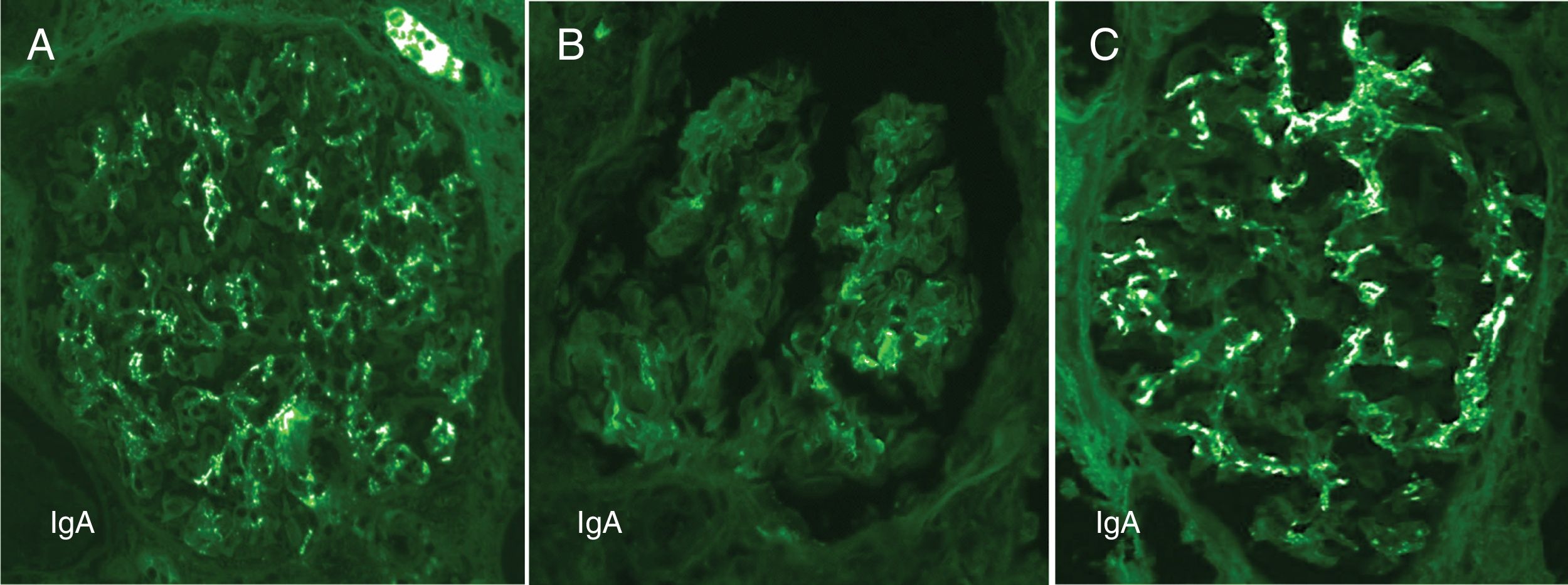
Contrasting features of IgA dominant IRGN and idiopathic IgAN.

Mesangial and capillary wall coarse granular deposits are prominent in exudative IgA IRGN (A) and less so in resolving IgA IRGN (B). These immunofluorescence patterns contrast with the largely mesangial deposits of IgA in idiopathic IgAN (C).

(0.23MB).
Fig. 2.

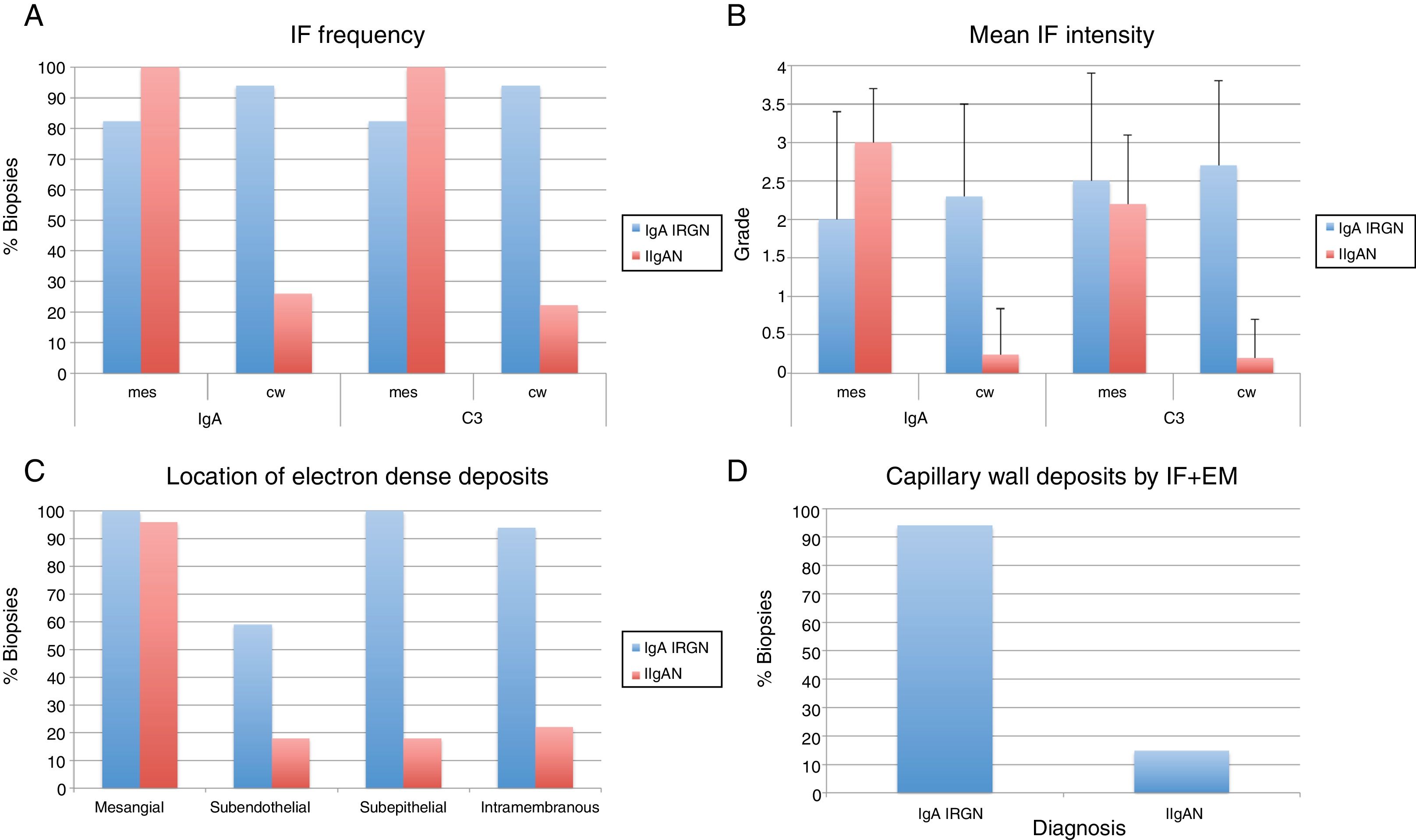
Analysis of immunofluorescence and electron microscopic features of IgA IRGN and idiopathic IgAN.

Mesangial deposits of IgA and C3 were seen with comparable frequency in IgA IRGN and idiopathic IgAN, however, there were more frequent (A) and intense (B) deposits of these reactants in the glomerular capillary walls (p<0.001 by Fisher Exact and Mann–Whitney tests respectively). Capillary wall deposits were more frequently observed by EM in IgA IRGN compared with idiopathic IgAN (C). Ninety-four percent of IgA IRGN had capillary wall deposits detectable by both IF and EM compared with 14.8% of idiopathic IgAN (D).

(0.27MB).

All patients were adults (mean age 55.5 years (range 30–93)), with acute kidney injury in 16 of 17 (mean serum creatinine 3.9±3mg/dL), proteinuria (4 in the nephrotic range) and hematuria. Seven patients had diabetes mellitus, 4 of whom had diabetic nephropathy. Twelve had known clinical features of recent or concurrent bacterial infection (involving skin 7, joint 1, lung 2, appendix 1, vascular graft 1), 6 of which had culture positive S. aureus. In 6 cases the organism is unknown to us, and 5 patients had limited available clinical data. Of 10 patients with available follow-up data, 6 developed end-stage kidney disease, 3 had partial recovery and 1 had complete recovery of renal function.

Acute or resolving IgA IRGN accounted for nearly 35% of renal biopsies with IgA deposits and 5.5% of all native renal biopsies received over the study period. For perspective, a review of 21 339 native kidney biopsies obtained in the United States and Canada over approximately 20 years, suggests a frequency of about 0.6% for IgA IRGN.1–4 Our observations show a 9-fold increase in frequency of IgA IRGN albeit in a small single practice sample over a short time span. IgA IRGN has distinctive features including strong C3 staining, capillary wall immune deposits and acute tubulointerstitial injury, but may be difficult to distinguish from idiopathic IgAN, a feature noted by others.5 IRGN may be a more significant differential diagnostic consideration in biopsies with predominant glomerular IgA deposition than has been generally recognized.

References
[1]
S.H. Nasr, G.S. Markowitz, J.D. Whelan, J.J. Albanese, R.M. Rosen, D.A. Fein, et al.
IgA-dominant acute poststaphylococcal glomerulonephritis complicating diabetic nephropathy.
Hum Pathol, 34 (2003), pp. 1235-1241
[2]
M. Haas, L.C. Racusen, S.M. Bagnasco.
IgA-dominant postinfectious glomerulonephritis: a report of 13 cases with common ultrastructural features.
Hum Pathol, 39 (2008), pp. 1309-1316
[3]
S. Worawichawong, L. Girard, K. Trpkov, J.C. Gough, D.B. Gregson, H. Benediktsson.
Immunoglobulin A-dominant postinfectious glomerulonephritis: frequent occurrence in nondiabetic patients with Staphylococcus aureus infection.
Hum Pathol, 42 (2011), pp. 279-284
[4]
A.A. Satoskar, S. Suleiman, I. Ayoub, J. Hemminger, S. Parikh, S.V. Brodsky, et al.
Staphylococcus infection-associated GN – spectrum of IgA staining and prevalence of ANCA in a single-center cohort.
Clin J Am Soc Nephrol, 12 (2017), pp. 39-49
[5]
A.A. Satoskar, G. Nadasdy, J.A. Plaza, D. Sedmak, G. Shidham, L. Hebert, et al.
Staphylococcus infection-associated glomerulonephritis mimicking IgA nephropathy.
Clin J Am Soc, 1 (2006), pp. 1179-1186
Copyright © 2019. Sociedad Española de Nefrología
Descargar PDF
Idiomas
Nefrología
Opciones de artículo
Herramientas