Dear Editor:
Crossed-fused renal ectopia is the second most common variety of renal fusion, with an incidence of 0.01% in the general population. There are at least six varieties of crossed-fused renal ectopia, and it is thought to be produced by a change in the migration of the kidney due to a vascular obstacle, or due to genetic or teratogenic factors. It is generally associated with other alterations of the gastrointestinal and locomotor systems.
Clinical case
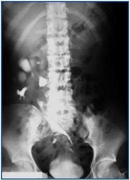
Female patient, aged 40 years, with a personal history of recurrent cystitis and two vaginal births, a habitual smoker of 10 cigarettes/day came to the Emergency Room due to pain in the right ileal fossa, fever and urination syndrome. An analysis was performed, which revealed leukocytosis with a left deviation (23,436 leukocytes/ul, 93% segmented), 456,000 platelets/ul, renal function within normal range (urea 34.1mg/dl and creatinine 0.9mg/dl), globular sedimentation velocity (GSV) of 42mm/hour within the first hour, Creactive protein at 34.1 and pyuria in the sediment (50 leukocytes per field), positive nitrite test and bacteria. At that point, a urine culture was extracted that came back positive for Escherichia coli 96 hours later. After extraction of the urine culture, an empirical antibiotic treatment was started with amoxicillin/clavulanic and gentamycine; treatment with both antibiotics was finished, since the antibiogram confirmed that the microorganism was sensitive to them. In the abdominal ultrasound, we see a structure compatible with crossed renal ectopia in the right ileum fossa, which is fused at its upper point with the right kidney, and no kidney in the left renal fossa. With the clinical, analytical and imaging data, the diagnosis of acute pyelonephritis with a crossed ectopic kidney was carried out. An anterograde pyelography was performed, in which both kidneys can be observed fused together and located in the right hemiabdomen (figure 1).
Discussion
Crossed ectopia without or with fusion (90% of all cases) occurs when the ectopic kidney is located on the side opposite to its ureter’s insertion point in the bladder. This is a rare congenital defect.1,2 At times, blood vessels responsible for irrigating the ectopic kidney cross the midline and can be responsible for stenosis of the pyelourethral union of the ectopic kidney or its normal counterpart.3 Most of these cases evolve asymptomatically and the diagnosis is made when a disease affects the ectopic kidney, such as an infection, lithiasis, tumours or other more infrequent possibilities.1,2,4 The diagnosis is performed by ultrasound, intravenous urography or anterograde pyelography,5 isotopic studies, computed tomography or nuclear magnetic resonance imaging. The treatment for this congenital defect merely concerns the pathology affecting the kidney; no other treatment is necessary if the patient is asymptomatic.
Figure 1.


