Dear Editor,
Studies suggest that incidence of calciphylaxis is 1% per year, with a prevalence of 4% among dialysis patients,1 however it is rarely present in kidney transplant patients or in those with stage 3 or 4 chronic kidney disease.2
The proximal distribution of lesions and the presence of ulceration are associated with a very poor prognosis, mainly because of wound infection and the subsequent death of the patient.3 In the cases of calciphylaxis described in kidney transplant patients, the prognosis may be even worse4,5 and the possible role of corticosteroids as a precipitant of the disease has also been discussed. However, although the pathogenesis of this condition is not well known, there are risk factors that could possibly contribute to proximal calciphylaxis and distal calciphylaxis in different ways. Therefore, no specific treatment has been established and in some cases a multidisciplinary and even empirical approach is needed. In any case, it is important to focus on normalising the phosphocalcic product and PTHi levels if they are elevated, since these are potential precipitants.6
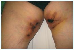
We would like to present the case of a 66-year-old female patient who underwent her first liver transplant 11 years ago because of chronic alcoholinduced liver disease. She underwent a second kidney transplant three years before this admission because of mesangial glomerulonephritis caused by IgA deposits, which presented hardened lesions with central dermal necrosis that were symmetrical and very painful, located on the inner thigh on both legs and that indicated calciphylaxis (figure 1). A cutaneous punch biopsy was carried out and the diagnosis was confirmed. However, a bone scan showed no extraskeletal uptake. Her usual treatment consisted of furosemide, bisoprolol, prednisone, tacrolimus, mycophenolate mofetil, omeprazole and acenocoumarol (since she presented chronic atrial fibrillation) and subcutaneous darbopoetin. In the tests carried out, the following results stood out: CRP 51mg/l, Hb 10g/dl and creatinine 2.9mg/dl, because of chronic nephropathy of the graft, with proteinuria 2.6g/day, cholestatic pattern with GGT 230U/l and alkaline phosphatase 177U/l, corrected calcium concentration 8.6mg/dl, phosphorous 6.6mg/dl and initial PTHi 653pg/ml. Treatment with cinacalcet 30mg/day and aluminium hydroxide used as a phosphorus binder was administered. Phospocalcic products were normalised and PTHi values were stabilised at 150pg/ml. Despite this, large ulcers developed and enzymatic ointment (Iruxol Mono®), and moist gauzes were applied locally on a daily basis. Opiate derivatives were administered orally for pain relief, as well as 50g intravenous sodium thiosulphate three times a week. Her clinical progress was not satisfactory and haemodialysis was necessary 38 days after diagnosis via a catheter in the right internal jugular vein because of deteriorated kidney function. At the same time, it was also necessary to maintain the correct plasma levels of tacrolimus and avoid any accompanying septic symptoms. A few hours later she suffered nonrecoverable cardiac arrest. No autopsy was carried out.
Despite our patient¿s fatal outcome, we would like to highlight the potential therapeutic benefits of cinacalcet in the treatment of proximal calciphylaxis with secondary hyperparathyroidism.7 Its usefulness in transplant patients with calciphylaxis is yet to be demonstrated, although its effectiveness in controlling hyperparathyroidism has already been described.8 Nor have we found any descriptions of other kidney transplant patients who were administered sodium thiosulphate, although its effectiveness has been demonstrated in several published studies involving dialysis patients. It has been observed that this drug is highly soluble in calcium thiosulphate form as it inhibits calcium precipitation and dissolves calcium deposits in tumours and calciphylaxis.9
We hope that by describing other cases of calciphylaxis in kidney transplant patients we are able to raise awareness about the use of treatments like cinacalcet, sodium thiosulphate and bisphosphonates, among others. Although, in this case, bone scan was uninformative, it seems that this procedure has a high sensitivity for diagnosing this disease, showing an abnormal isotope uptake on a subcutaneous level in 97% of cases.10
Figure 1.


