To the Editor:
High blood pressure (HBP) occurs in 1% of children,1 and 10% of cases are renovascular in origin; fibromuscular dysplasia (FD), Takayasu arteritis (TA) and the middle aortic syndrome (MAS) are the most commonly associated aetiologies. These diseases pose difficulties in differential diagnosis due to their clinical similarities. This article describes the case of a girl with MAS as aetiology of HBP. Reports on this disease in paediatric age are scarce, as was its form of presentation and evolution.
CASE STUDY
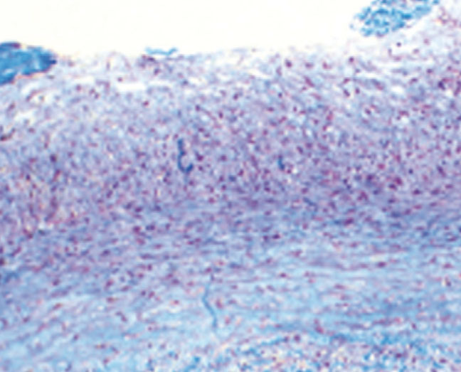
Patient who was hospitalised when she was two years due to HTA associated with loss of strength and sensation in her lower limbs. The arteriography showed decrease in aortic diameter, 20% stenosis at the right renal ostium, critical stenosis of the left renal artery and no flow in the left lower renal pole. The decision was made to perform left renal autotransplantation with renal anastomosis of the iliac artery and biopsy of the renal artery, which reported findings consistent with FD (Figure 1). She was discharged with minoxidil and propranolol.
A year later, the patient is hospitalised due to hypertensive crisis. The renal scintigraphy showed exclusion of the left autotransplanted kidney and impaired perfusion of the right kidney. Due to suspected large vessel vasculitis type TA, paediatric rheumatology assessment was requested. Although the patient met the criteria for TA classification (abdominal aorta and renal arteries stenosis associated with HBP), prior biopsy showed no findings of vasculitis.
Further differential diagnoses pointed to FD but this was ruled out because FD renal lesions have a characteristic image of pearls necklace2 and rarely affect the ostium or the proximal segments. In this patient, there was no image of pearls necklace and the involvement of the renal artery was at the proximal portion of the artery. Following these imaging findings, MAS was eventually diagnosed.
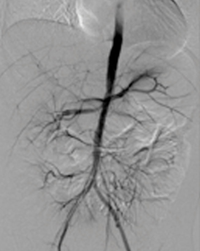
Another arteriography was performed, which reported irregular abdominal aorta with progressive distal thinning, arterial anastomosis autotransplant occlusion and progression of right renal artery stenosis (Figure 2); it was decided that a right renal primary angioplasty was to be carried out, which was unsuccessful due to the persistence of stenosis. Because of the difficult monitoring of blood pressure figures, selective venous sampling of the renal veins was performed to measure renin, with a difference of 10:1 being found in the concentrations of autotransplanted kidney against the right kidney. This confirmed the suspicion of renovascular HBP originating in the autotransplanted kidney. It was not possible to perform autotransplanted renal artery embolisation due to the risk of extensive necrosis, as parasitic branches were found that provided flow to the autotransplanted kidney and the intrinsic muscles of the pelvis. It was decided to carry out nephrectomy of the left autotransplant. Evolution was satisfactory with better control of blood pressure figures, and as such, the patient was discharged.
DISCUSSION
Renovascular hypertension is defined as the presence of HBP secondary to an obstructive lesion of the renal arteries3; it is the third leading cause of HTA4 amongst children and major diseases that are associated with it are FD, TA and type 1 neurofibromatosis. MAS is a rare cause, but must be considered as a differential diagnosis.5
MAS is characterised by stenosis of the proximal abdominal aorta and ostial stenosis of its major branches (renal arteries 90%)2; the mean age of onset is 20 years,4 although recent papers report earlier diagnosis.6,7 Its aetiology is diverse; most cases are idiopathic,4 although it has been associated with diseases such as neurocutaneous syndrome and Williams syndrome.6 Among the hypotheses proposed is inadequate fusion of the two dorsal aortas during embryonic development and it has also been linked to congenital rubella syndrome.8 The most important clinical marker is severe HBP caused by ostial stenosis of the renal artery, which leads to increased production of renin,4 as happened in this patient. Commonly observed histological findings are intimal fibrodysplasia, distortion of internal elastic lamina with absence of inflammatory changes, with lesions similar to those observed in some forms of FD.9
For its diagnosis, arteriography remains the gold standard; it is also useful to define the extention of disease and conduct surgical therapy.10 Additional tests are measuring renin levels and renal function tests. In differential diagnosis, it is important to note that the acute phase reactants are normal, unlike with TA, and there are no systemic symptoms such as fever, weight loss, abdominal pain or skin lesions.
Treatments vary depending on the severity of each case, and range from conservative to surgical management, such as autotransplant6 and nephrectomy when revascularization is not possible.4,7 Percutaneous transluminal balloon angioplasty and stenting are used in older children,10 however, successful results have not been displayed, as is the case with this patient.10 Medical therapy is important for aggressive control of blood pressure, which is often difficult to manage.8
To conclude, this article reports the case of a girl diagnosed with middle aortic syndrome caused by renovascular hypertension, with a clinical presentation that proved to be a diagnostic challenge, as it was difficult to distinguish from other more common causes, such as TA and FD. Associated HBP was difficult to treat and required multiple therapeutic interventions such as autotransplant and balloon angioplasty, which were unsuccessful and required nephrectomy for adequate control of blood pressure.
Conflicts of interest
The authors declare that they have no conflicts of interest related to the contents of this article.
Figure 1. Renal artery biopsy
Figure 2. Abdominal arteriography


