Dear Editor,
In connection with the clinical case presented in number 4, volume 26, of this same journal, we would like to report a similar case of intussusception of the terminal ileum by a carcinoid tumour in a patient with chronic kidney failure as it is an infrequent and little referenced illness in patients with chronic kidney failure.1
A54 year old female patient with a history of chronic renal failure, hyperuricaemia and nephrolithiasis. She attended Accident and Emergency with generalized abdominal pain, nausea, vomiting and diarrhoea of 48 hour duration.
She had distended and tympanic abdomen with diffuse pain and no signs of peritonism.
Air-fluid levels could be seen in the small intestine on plain abdominal x-ray. The CT of the abdomen showed a dilated jejunumileum with thickening of the terminal ileum and caecum wall and a 4cm mass.
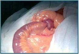
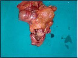
With the impression of an acute intestinal obstruction, the patient underwent urgent laparotomy. The small intestine was dilated to the terminal ileum where a tumour measuring 5cm was found that had caused the intussusception of the small intestine and the obstruction. A right colectomy and an ileum-colonic anastomosis were performed.
The postoperative evolution was satisfactory. A carcinoid tumour of the ileum measuring 1.8 x 1.5cm was infiltrating the adjacent muscle on anatomo-pathological examination; the tumour was serotonin-producing.
The most frequent location of the intestinal carcinoid tumours (ICT) is the caecal appendix (50%), followed by the ileum (25%), as in our case. The symptoms usually appear late and in a non-specific manner leading to a late diagnosis. 60% of the patients present hepatic metastases at the time of diagnosis. The carcinoid syndrome is found in only 5% of the patients and is related to the presence of hepatic metastases.2
In the diagnosis,3 aside from the conventional imaging tests, the Octroscan is useful as it provides information on the localisation of tumours larger than 0.5cm and the expression or not of serotonin receptors. The differential diagnosis includes other intestinal tumours, ileocaecal Crohn’s disease and systemic mastocytosis.
Treatment should include surgical removal provided there are no metastases. Epsilon-aminocaproic Acid or somatostatin should be administered during the intervention to avoid carcinoid crisis. When there is metastatic dissemination, the patient should be managed conservatively. To reduce the metastases, 5-Fluorouracil or Adriamycin have been combined with Streptozotocin. Different drugs have been used for the treatment of the carcinoid syndrome with varied results including Chlorophenylalanine, serotonin antagonists, somatostatin, octeotride and interferon.
Figure 1.
Figure 2.


