Dear Editor,
Hypertension in the elderly is mainly essential with coarctation of the aorta a secondary cause.1 The median survival of patients with coarctation of the aorta is low: only 25% live past 50 years of age.2,3 Most cases are women, due to a lower tendency to develop atherosclerosis and hypertension.4
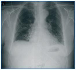
Below is the case of a male patient aged 83 who was admitted for surgery of the left paranasal squamous cell carcinoma, which appeared as a complication in a compressive cervical haematoma that required urgent tracheotomy. His background revealed longstanding refractory hypertension. A physical examination revealed a normal cardiopulmonary auscultation with distal pulses present on the upper limbs and diminished in the lower. Blood pressure in the upper right extremity was 182/81mmHg, significantly higher than the left side, where it was 130/75mmHg. The latter was similar to those of the lower extremities. Analytically, the data showed no renal secondary hypertension, thyroid or kidney disease. The echocardiogram revealed a significant hypertrophy of the left ventricle in septal location. A slight cardiomegaly and the inverted E sign (Figure 1) were observed on the radiograph which, together with the difference in blood pressure in both upper extremities, directed us towards a diagnosis of probable aortic coarctation.
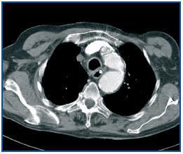
Therefore, a chest CT was performed with contrast. This revealed, in the aortic arch, distal to the supra-aortic trunks, a poststenotic dilatation of a maximum of 3.7cm in diameter, compatible with aortic coarctation (Figure 2).
After assessing the clinical status, the tumour staging (T2, N2b, M0) and the high comorbidity of surgery, conservative treatment was chosen.
In conclusion, the diagnosis of aortic coarctation should always be discarded for any patient with refractory hypertension. A proper physical examination with palpation of distal pulses and measurement of blood pressure control between extremities is a good guide towards diagnosis.
Figure 1.
Figure 2.


