To the Editor:
Acquired perforating dermatosis (APD) is an uncommon cutaneous perforating disorder. It is characterised by the presence of hyperkeratotic papules and nodules and histologically, by transepidermal elimination of various substances, such as keratin, collagen and elastic fibres. It was first described by Mehregan in 1967. There are two types: hereditary and acquired, the latter is generally associated with diabetes mellitus (DM) and chronic renal failure (CRF).1
In the literature, we also found other cases associated with neoplasia,2,3 liver diseases and endocrine disorders,4 AIDS,5 tuberculosis,6 etc.
There are four classic forms of APD, according to clinical and histological criteria: elastosis perforans serpiginosa, reactive perforating collagenosis, perforating folliculitis and Kyrle disease.
As regards the population with CRF on renal replacement therapy, the only studies of this condition date back to 1982,7 with a reported prevalence of 4.5%-10% in haemodialysis patients in the United States, and 1996,8 with a prevalence of 11% in the United Kingdom. Only haemodialysis patients have been reported to date, not peritoneal dialysis patients.
Many treatments have been used in an attempt to manage this disorder, with varying results: topical and systemic steroids, topical and systemic retinoids, vitamin A, light therapy, methotrexate and cryotherapy. In recent times, allopurinol has been considered as treatment, due to its antioxidant effect: inhibiting xanthine oxidase and reducing the production of oxygen free radicals and, therefore, collagen damage.9,10
We report two cases in our unit; the first was a peritoneal dialysis patient and the second, a haemodialysis patient.
CASE 1
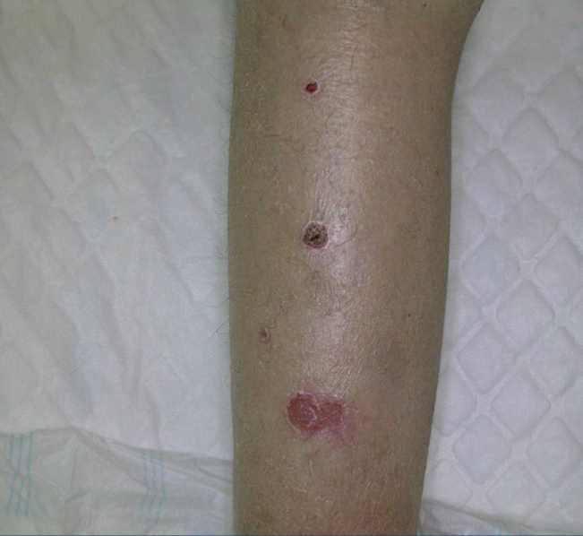
A 39-year-old female with a history of very debilitating juvenile chronic arthritis from the age of 16 years old. She was not diabetic and had bilateral hip prostheses due to femoral head avascular necrosis, secondary to prolonged steroid treatment. She also had rheumatic mitral regurgitation and was on peritoneal dialysis due to chronic kidney disease. She came to our hospital due to exophytic and hyperkeratotic lesions on the soles of her feet, the pretibial area (Figure 1) and her buttocks. She was transferred to the Dermatology department, the lesions were biopsied and she was diagnosed with reactive perforating collagenosis. After reviewing the literature, treatment was started with allopurinol and a significant improvement was noted in the lesions after one month, with the disappearance of pruritus.
CASE 2
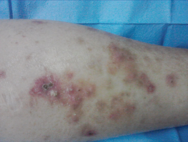
A 50-year-old male with ESRD secondary to diabetic nephropathy on renal replacement therapy with haemodialysis. He had chronic ischaemic heart disease, ischaemic dilated cardiomyopathy, diabetic retinopathy and had suffered a stroke some years before; he was admitted to internal medicine due to an episode of diabetic ketoacidosis. While hospitalised, multiple maculopapular erythematous lesions were detected with universally distributed central hyperkeratotic scaly areas (Figure 2). We consulted the Dermatology department, a skin biopsy was taken of the lesions and the patient was diagnosed with Kyrle disease.
DISCUSSION
Perforating skin disorders are characterised by the transepidermal elimination of some components of the dermis and they have classically been classified into four types: elastosis perforans serpiginosa, reactive perforating collagenosis, perforating folliculitis and Kyrle disease.
These four disorders have been reported in patients with CRF, DM or both conditions. APD lesions associated with CRF or DM are usually 2-10mm, hyperkeratotic and are often umbilicated papules generally located in the limbs, particularly the legs. The lesions are usually very pruritic, with positive Koebner phenomenon on scratching. In the cases that we reported, there was a predominance of lower limb lesions and in both cases, pruritus was the main symptom. The presence of lesions on the face, hands and feet is exceptional. Our second patient had major lesions in both feet.
APD was also reported in other cases of CRF not due to DM, including obstructive nephropathies, hypertensive nephroangiosclerosis, AIDS, etc. This suggests that the cutaneous changes typical of CRF may act as a trigger in the development of APD. Microdeposits of substances such as calcium salts may promote a local inflammatory reaction, as well as connective tissue degradation. In fact, microcrystal deposits have been observed in the upper dermis in ultrastructure studies carried out on patients with APD.
In conclusion, this pathology is relatively common in dialysis units (prevalence varies between 4% and 10%). It is not always diagnosed and it is occasionally debilitating, due to the pruritus that it causes. Its pathogenesis is unknown, although it may be influenced by the trauma caused by pruritus itself. In any case, it is a relatively unknown condition, and as such, new studies are necessary in order to better define this collagen abnormality.
Conflicts of interest
The authors declare that they have no conflicts of interest related to the contents of this article.
Figure 1. Pretibial lesion.
Figure 2. Universally distributed hyperkeratotic and erythematous lesions.


