


ANCA-associated vasculitis (AAV) is a rare autoimmune disease that causes inflammation of small blood vessels, often leading to organ damage, particularly in the kidneys. Biomarkers like calprotectin and CD163 have been proposed as indicators of disease activity and inflammation. This study aims to analyze the dynamics of calprotectin and usCD163 during stable remission.
MethodsThis retrospective observational study included AAV patients in remission receiving rituximab as maintenance therapy. Seventeen patients were included, with 78 samples available for analysis. Serum calprotectin and usCD163 levels were measured using ELISA, and clinical data were collected from electronic medical records.
ResultsSeventeen AAV patients in remission were analyzed. usCD163 and serum calprotectin levels showed a significant positive correlation (p=0.005). Both biomarkers correlated with CRP, and usCD163 also correlated with ESR. Levels of both biomarkers were higher within 6 months after a disease flare. During follow-up, mean biomarker levels were higher in patients who later relapsed, though only usCD163 reached statistical significance (p=0.05). Calprotectin levels were significantly higher in patients who had received rituximab less than three months prior compared to those treated 9–12 months earlier (p=0.031).
ConclusionssuCD163 and serum calprotectin may serve as biomarkers for disease activity and relapse in AAV. Their correlation with renal function outcomes and higher levels in patients who relapsed suggest that they might be useful to measure persistent immune activity. Further studies are needed to explore their role in predicting flares and guiding personalized treatment strategies.
La vasculitis asociada a ANCA (AAV) es una enfermedad autoinmune rara que causa inflamación de los pequeños vasos sanguíneos, lo que a menudo conduce a daño orgánico, especialmente en los riñones. Se han propuesto biomarcadores como la calprotectina y CD163 como indicadores de actividad de la enfermedad e inflamación. Este estudio tiene como objetivo analizar la dinámica de la calprotectina y usCD163 durante la remisión estable.
MétodosEste estudio observacional retrospectivo incluyó a pacientes con AAV en remisión que recibían rituximab como terapia de mantenimiento. Se incluyeron 17pacientes, con 78 muestras disponibles para análisis. Los niveles de calprotectina sérica y usCD163 se midieron mediante ELISA, y los datos clínicos fueron recopilados de los registros médicos electrónicos.
ResultadosSe analizaron 17 pacientes con AAV en remisión. Los niveles de usCD163 y calprotectina sérica mostraron una correlación positiva significativa (p=0,005). Ambos biomarcadores se correlacionaron con la proteína C reactiva (CRP), y usCD163 también se correlacionó con la velocidad de sedimentación globular (ESR). Los niveles de ambos biomarcadores fueron más altos dentro de los 6meses posteriores a un brote de la enfermedad. Durante el seguimiento, los niveles medios de biomarcadores fueron más altos en los pacientes que posteriormente presentaron una recaída, aunque solo usCD163 alcanzó significación estadística (p=0,05). Los niveles de calprotectina fueron significativamente más altos en los pacientes que habían recibido rituximab menos de 3meses antes en comparación con aquellos tratados entre 9 y 12meses antes (p=0,031).
ConclusionesusCD163 y la calprotectina sérica pueden servir como biomarcadores de actividad de la enfermedad y recaída en la AAV. Su correlación con los parámetros de función renal y sus niveles elevados en pacientes que presentaron recaída sugieren que podrían ser útiles para medir la actividad inmune persistente. Se requieren más estudios para explorar su papel en la predicción de brotes y en la personalización de estrategias de tratamiento.
Anti-neutrophil cytoplasmic antibody (ANCA)-associated vasculitis (AAV) is a multisystemic disease that causes inflammation of small blood vessels, often affecting the kidneys. The disease is triggered by ANCA binding to proteins like proteinase 3 (PR3) and myeloperoxidase (MPO) on neutrophils, which leads to neutrophil degranulation and the release of inflammatory molecules.1 Treatment involves intensive immunosuppressive therapy during the induction phase, followed by a maintenance phase. Anti-CD20 targeted therapy with rituximab has demonstrated its efficacy both in the induction to remission phase as in the maintenance phase.2 Rituximab not only abrogates ANCA production but also inhibits neutrophil responses, although the exact mechanism remains unclear. Some researchers have proposed that this may be associated with changes in stromal-derived factor-1 (SDF-1) levels during B-cell recovery, potentially affecting neutrophil egress from the bone marrow. Others have suggested alternative mechanisms, including neutropenia caused by autoantibody production following rituximab treatment and the expansion of large granular lymphocytes that may induce neutrophil apoptosis.3 While guidelines suggest a maintenance period of at least 24 months, the optimal duration remains unclear. In this setting, biomarkers to assess disease activity, predict relapses, and tailor treatment plans accordingly are a topic of interest.
Calprotectin is a protein released by activated monocytes and neutrophils that promotes the inflammatory response in AAV leading to increased transcription of proinflammatory cytokines and facilitating leukocyte recruitment to inflammation sites.4 Due to its low molecular weight (36.5kDa), calprotectin diffuses into the bloodstream, where it can be measured, though it has a short half-life of only 5h.5 Some authors have observed higher serum calprotectin concentration in AAV patients in the acute phase of the disease compared to patients in remission.6–8 Moreover, Pepper et al. found that, in PR3-ANCA patients in the acute phase treated with rituximab, and increase in serum calprotectin by month 2 or 6 compared to the moment of the diagnostic was associated to a higher risk of relapse at 18 months.9 On the contrary, the dynamics of calprotectin levels during remission have been less extensively studied. Our group examined the evolution of serum calprotectin levels in a cohort of patients in stable remission over a 2-year period.6 Additionally, we observed higher calprotectin levels in patients who showed a greater decline in glomerular filtration rate (GFR) and worsening proteinuria during the 2-year follow-up in remission, but we were unable to correlate calprotectin levels with an increased risk of relapse. Romand et al., in a post-hoc analysis of the MAINTRITSAN study, demonstrated that patients in remission who showed increasing calprotectin levels during the first 6 months after initiating maintenance treatment had worse renal outcomes during follow-up and a tendency toward a higher risk of AAV relapse.10 In both studies, the maintenance treatment was not homogenous and included rituximab in different schedules of administration or the administration of antiproliferative drugs such as azathioprine or mycophenolate, together with low-dose steroids.
CD163 is a membrane protein found on the surface of monocytes and macrophages. The soluble form of CD163 (sCD163) is generated through enzymatic cleavage during inflammation and is associated with excessive M2 macrophage activation.11 O’Reilly et al. reported that urinary sCD163, shed by macrophages, correlates with active glomerular inflammation in AAV.12 B-cell depletion by rituximab is mediated by various immune mechanisms, including the phagocytosis of rituximab-opsonized B cells by M2 macrophages.13 In a cohort of 24 incident AAV patients, Villacorta et al. observed a decline in usCD163 levels during the first year after diagnosis in all patients who responded to treatment and reached remission.11 In contrast, 5 patients who relapsed during follow-up showed a significant increase in usCD163 compared to previous levels. Although some authors have reported significantly lower usCD163 levels in remission compared to active AAV, the dynamics of CD163 during stable remission have not been studied.
This study aims to comprehensively describe the dynamics of two recognized biomarkers of AAV activity – calprotectin and usCD163 – during periods of stable remission. Additionally, it seeks to explore the correlation between these biomarkers and with other markers of inflammation and disease activity, their relationship with relapses, and with rituximab administration.
MethodsStudy populationThis is a retrospective observational study. All patients were diagnosed with AAV according to the Chapel Hill Consensus Criteria: compatible clinical symptoms, ANCA positivity, and a kidney biopsy indicative of AAV, and were in remission of the disease at the moment of the inclusion. The Birmingham Vasculitis Activity Score (BVAS) was used to determine disease activity, and patients in remission were defined as having a BVAS of 0. All patients were receiving rituximab as maintenance treatment. Remission had previously been achieved with intravenous methylprednisolone boluses followed by cyclophosphamide or rituximab, as recommended by the EULAR/ERA-EDTA guidelines.
The frequency of rituximab re-administration during remission in our cohort was guided by CD20+ lymphocyte repopulation and ANCA titer kinetics. The dose administered was 500mg. In the MAINRITSAN2 study,14 a randomized controlled clinical trial, fixed-interval re-administration of 500mg rituximab every 6 months was compared with a personalized regimen: re-administration at the time of CD20+ lymphocyte repopulation or ANCA re-positivation (or a significant increase compared to baseline). The study found no significant differences in relapse rates between the two groups; however, the number of rituximab infusions was lower in the personalized regimen group.
Contraindications to participate in the study were defined as: active neoplasm or infection, other autoimmune diseases and end-stage kidney disease (ESKD) requiring renal replacement therapy.
Samples included in this study were provided by the HUB-ICO-IDIBELL Biobank (PT20/00171). Biologic products were processed according to established procedures.
This study was approved by the Bellvitge University Hospital Review Board.
Sampling and biomarker assaysPrior to each routine clinical visit at the outpatient clinic, lab tests were performed, and serum and urine samples were obtained for biomarker assays. We determined serum calprotectin and usCD163 using a commercial ELISA kit (Legend Max Human MRP8/14, Biolegend Inc.; for calprotectin, and the Quantikine® Human CD163 immunoassay, Bio-Techne R&D Systems, for CD163) according to the manufacturer's instructions.
Clinical dataClinical and analytical data for the patients were obtained from electronic medical records. We recorded sex, age, ANCA specificity (MPO/PR3) and titers, the presence of hematuria and proteinuria, CRP values, serum creatinine and GFR. We collected analytical data at the time of diagnosis, recruitment, and successive clinical visits. Histology at diagnosis was analyzed according to Berden's classification. The immunosuppressive treatment, as well as patient and renal survival, were also recorded.
Statistical analysisData analysis was performed using IBM SPSS Statistics Version 26.0 (IBM Corp., Armonk, NY, USA) and GraphPad Prism version 8.00 (GraphPad Software, La Jolla, CA, USA). Continuous variables were expressed as mean±standard deviation (SD) and categorical variables as total number (n) and percentage (%). For comparison between quantitative and normally or not-normally distributed variables, Student's t-test and Mann–Whitney U were performed respectively. Chi-square test was used for qualitative variables. Spearman's correlation was applied to correlate not normally distributed variables. p-Values were two-tailed and statistical significance level was established at p<0.05.
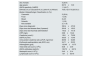
ResultsBaseline characteristicsTable 1 shows the main baseline characteristics of the populations at the time of recruitment. Seventeen patients diagnosed with AAV in remission and undergoing maintenance treatment with rituximab were included.
The main baseline characteristics of the patients.
| Sex (%male) | 5 (29.4) |
| Age (years) | 66.73±10.9 |
| ANCA specificity (%MPO) | 11 (64.7) |
| Ethnicity (% (n) Caucasian/% (n) Latino/% (n) African) | 76.6 (13)/17.6 (3)/5.8 (1) |
| Berden Histopathologic Classification (n (%)) | |
| Crescentic | 5 (29.4) |
| Focal | 1 (5.9) |
| Mixed | 8 (47) |
| Sclerotic | 1 (5.9) |
| Not available | 2 (11.8) |
| Days since diagnostic | 390.4±472.8 |
| Days since last disease flare (if present) | 204.6±109.2 |
| Days since last rituximab administration | 143.2±101.8 |
| CRP (mg/L) | 6.88±10.42 |
| Creatinine (μmol/L) | 158.1±79.28 |
| eGFR (mL/min) | 38.35±15.46 |
| Urine protein creatinine ratio (uPCR, mg/mmol) | 88.56±94.86 |
| Erythrocyte sedimentation rate (ESR, mm) | 34.71±23.33 |
| Haemoglobin (g/dL) | 123.40±13.73 |
| Total white cell count (×109/L) | 8.38±2.49 |
| ANCA antibodies (karbU/L) | 106.70±247.6 |
| Neutrophil count (×109/L) | 5.40±21.4 |
| Lymphocyte count (×109/L) | 1.91±6.619 |
ANCA, anti-neutrophil citoplasmic antibody; MPO, myeloperoxidase; CRP, C-reactive protein; eGFR, estimated glomerular filtration rate.
During follow-up, a total of 73 samples of serum and urine were obtained from the patients recruited into the study to determine usCD163 and serum calprotectin levels.
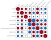
usCD163 and serum calprotectin levels showed a significant positive correlation (r=0.361, p=0.005). When exploring the correlation of these biomarkers with other inflammation markers, both biomarkers were significantly correlated with serum C-reactive protein (CRP), showing a positive correlation, and usCD163 was also positively correlated with erythrocyte sedimentation rate (ESR). Additionally, both biomarkers negatively correlated with the number of circulating lymphocytes. ANCA titre did not correlate with any of the biomarkers. Interestingly, only serum calprotectin levels correlated with kidney function parameters (see Fig. 1).
Dynamics of suCD163 and serum calprotectin in relation to disease activity
As expected, the time in remission after the last recorded disease activity influenced the levels of the biomarkers. Both usCD163 and serum calprotectin were higher in samples obtained within 6 months after a disease flare compared to those obtained more than 6 months later (Fig. 2A). We also analyzed the levels of both biomarkers according to the time elapsed since the last disease activity. A decrease in biomarker levels was observed during the first year compared to the levels observed within the first 3 months, after which they remained stable (Fig. 2B).

Illustrates biomarker levels based on the time elapsed since the last recorded activity. (A) Biomarker levels were higher in patients whose last disease relapse occurred less than 6 months ago compared to those with a relapse more than 6 months ago. (B) A decrease in biomarker levels was observed during the first year compared to the levels observed within the first 3 months, after which they remained stable.
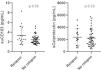
During follow-up, 4 patients experienced a relapse. Nineteen samples obtained during remission prior to relapse were available from these 4 patients (ranging from 67 to 483 days prior to relapse). Table 2 shows the baseline analytical data of the patients at the time of recruitment, categorized according to the subsequent development of relapse. Table 3 also details the main baseline characteristics of the patients who experienced a relapse during follow-up, as well as the evolution of their biomarkers. As detailed in Table 2, we did not find differences in ANCA titer, CRP levels, eGFR or proteinuria between patients who later relapsed compared to those who did not. We then compared the levels of the biomarkers in the samples obtained in remission from patients who later relapsed with the levels in samples from patients who did not relapse. During the follow-up period, the mean levels of serum calprotectin and usCD163 in remission were higher in patients who relapsed compared to those who did not. However, the differences reached statistical significance only for usCD163 (p=0.05) (Fig. 3).
The main baseline analytical parameters of the patients who relapsed during follow-up compared to the patients who did not relapse.
| Relapse | No relapse | p-Value | |
|---|---|---|---|
| ANCA titer (karbU/L) | 99±185.4 | 109.1±270.4 | 0.31 |
| Lymphocyte count (×106 cells) | 1.85±1.06 | 1.92±0.55 | 0.84 |
| sCreatinine (μmol/L) | 127±51.29 | 167.6±86.79 | 0.61 |
| eGFR (mL/min) | 42.75±12.12 | 37±16.54 | 0.53 |
| uPCR (g/mol) | 140.8±124.8 | 71.17±81.88 | 0.38 |
| CRP (mg/L) | 3.33±2.08 | 7.69±11.46 | 0.82 |
| suCD163 (ng/mL) | 2.58, IQR [2.16–4.57] | 2.19, IQR [1.77–2.86] | 0.05 |
| Calprotectin (ng/mL) | 2016, IQR [974.7–3336] | 1871, IQR [1210–2952] | 0.23 |
ANCA, anti neutrophil cytoplasmic antibody; sCreatinine, serum creatinine; eGFR, estimated glomerular filtration rate; uPCR, urinary protein to creatinine ratio; suCD163, soluble urinary CD163.
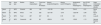
The baseline characteristics at diagnosis of patients who later experienced a relapse during follow-up.
| Sex, age | Anca type | Berden | Time to relapse(days) | Time since last RTX(days) | Absolut Δcalprotectin(pg/mL) | Absolut ΔusCD163(pg/mL) | Δcalprotectin prior to relapse(pg/mL) | Δus CD163 prior to relapse(pg/mL) | Organ involvement of relapse (BVAS) | |
|---|---|---|---|---|---|---|---|---|---|---|
| Patient 1 | 77, M | MPO | Sclerotic | 486 | 180 | −1431.58 | −2.89 | 734.42 | −0.21 | Renal (12) |
| Patient 2 | 63, F | MPO | Mixed | 179 | 262 | −2217.69 | −2.48 | −2227.69 | −2.48 | Renal (19) |
| Patient 3 | 79, F | MPO | Focal | 287 | 267 | NA | 0.78 | NA | 0.78 | Lung (6) |
| Patient 4 | 57, F | MPO | Crescentic | 481 | 98 | −259.04 | −1.03 | −33.88 | −1.13 | General features (2) |
Absolute Δ refers to the change in the biomarker between the first available sample taken during remission and the last available sample before relapse. Δ prior to relapse refers to the change in the biomarker between the two most recent samples taken prior to relapse. Time since last RTX refers to the duration between the last administered dose of rituximab and the occurrence of relapse.
M, male; F, female; RTX, rituximab; usCD163, urinary soluble CD163; NA, not available; BVAS, Birmingham vasculitis activity score.

We therefore investigated the influence of rituximab administration on the dynamics of biomarkers over time.
In our study, patients were followed from recruitment for an average of 306.81±174.10 days until the end of the follow-up period. During this time, they received an average of 1.5±0.65 doses of rituximab. From the initiation of remission maintenance therapy with rituximab until the end of the follow-up period, these same patients received an average of 2.68±1.01 doses, with a mean interval of 306±158.11 days between doses.
Initially, we observed a statistically significant negative correlation between serum calprotectin levels and the number of days elapsed since the last dose of rituximab (r=−0.359, p=0.004). In our cohort of patients treated with rituximab, calprotectin levels were higher in samples from patients who had received their last dose within the past 3 months compared to those whose last dose was 6–9 months or 9–12 months ago (p=0.028 and p=0.001, respectively). The kinetics of suCD163 showed a similar pattern, with samples obtained less than 3 months after rituximab administration exhibiting significantly higher levels compared to those obtained 3–6 months and 6–9 months after (p=0.034 and p=0.007, respectively).
To minimize the influence of disease activity on biomarker levels, we selected only samples in remission obtained at least six months after active disease. Fifty-two samples fulfilled this criterion. Calprotectin levels in patients who had received rituximab less than three months prior were significantly higher compared to those who had received rituximab 9–12 months earlier (p=0.031).). In contrast, we did not observe any changes in suCD163 levels based on the time elapsed since the last rituximab administration.
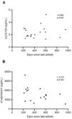
Finally, to assess the influence of rituximab, we selected remission samples obtained at least six months after active disease and at least six months after the last RTX dose. Twelve patients had 26 available samples, with a median remission duration of 365 days. For CD163, we observed a progressive increase over the follow-up period (correlation suCD163 vs days elapsed since last rituximab administration r=0.468, p=0.037). In contrast, calprotectin showed a decreasing trend during follow-up (r=−0.151, p=0.563) (see Fig. 4).

For comparison, we recruited six patients in remission who had previously received mycophenolate mofetil as maintenance therapy and were currently off treatment. We obtained 27 samples over a median follow-up of 151 days (IQR 35–245 days). Baseline characteristics at recruitment are shown in Supplementary Table 1. In contrast to patients in remission previously treated with rituximab, these patients exhibited stable levels of suCD163 and serum calprotectin throughout follow-up.
DiscussionAAV is a multisystemic disease characterized by inflammation of small vessels, which often affects the kidneys. Treatment consists of immunosuppressive therapy, which is more intense during the induction phase and is subsequently reduced in the maintenance phase of disease remission. The optimal duration of maintenance treatment has not been established by randomized controlled trials, although clinical guidelines recommend at least 24 months. Given the chronic, relapsing nature of AAV, several researchers have sought to identify biomarkers that can detect subclinical disease activity and predict future relapses during the remission phase, with the goal of individualizing the treatment schedule. The kinetics of several biomarkers have been extensively studied in the acute phase and early remission phase, but little is known about long-term remission. This latter phase is of the utmost importance, as it is when the clinician must decide whether to maintain or withdraw immunosuppressive treatment. Our findings provide new insights into the behavior of these biomarkers over time, their association with systemic inflammation and kidney function, and their potential to anticipate relapse. We also investigated how immunosuppressive maintenance therapy, particularly rituximab, may influence their dynamics.
Our first objective was to assess the correlation between serum calprotectin and usCD163 during remission. We found a statistically significant positive correlation between both biomarkers, suggesting that while they may reflect overlapping inflammatory pathways, they likely represent distinct immune processes. This is consistent with previous literature reporting that calprotectin is a marker of neutrophil and monocyte activation,15 whereas usCD163 reflects macrophage activity in renal tissue and urinary compartments.16 The concept of combining biomarkers that reflect different inflammatory pathways is not new and has been explored in previous studies aiming to improve diagnostic or prognostic accuracy. In the context of AAV, where disease activity can be heterogeneous and driven by multiple immune mechanisms, using a multimarker approach may enhance the performance of individual biomarkers. Several publications, from our group and other authors, have already proposed combining biomarkers such as calprotectin, CD163, and others to capture distinct aspects of disease pathophysiology, with the goal of achieving better sensitivity and specificity in detecting subclinical activity or predicting relapse.7,17,18 Thus, the fact that both biomarkers show a positive correlation supports their combined use – not only during active disease but also during remission – where they may capture complementary aspects of subclinical inflammation.
Secondly, these biomarkers showed consistency in correlating with other markers of activity, such as CRP,19 lymphocyte count20 and ESR. Importantly, we observed that only calprotectin correlated with kidney function parameters, highlighting its possible dual role in reflecting systemic inflammation and renal involvement.
A key goal of our study was to investigate whether elevated levels of these biomarkers during remission were associated with subsequent relapse. In our cohort, patients who relapsed had higher levels of both biomarkers during remission, although only usCD163 reached statistical significance. Most existing studies on usCD163 have focused on its potential as a biomarker for early detection of disease relapse, investigating its rise shortly before renal flares. In one prospective study, only a single patient exhibited an elevation in usCD163 levels prior to the clinical onset of relapse,11 suggesting limited predictive value based on current evidence. In contrast, our study specifically examines the dynamics of usCD163 during periods of stable remission, an aspect that has been less thoroughly explored. We observed that patients who eventually experienced a relapse had slightly higher usCD163 levels already during remission, compared to those who remained in sustained remission. However, these levels still fell within the low range typically associated with remission. This finding suggests that subtle variations in usCD163 expression during remission may be associated with an increased risk of future relapse.
Our group previously investigated serum calprotectin levels in AAV patients in remission and found that higher levels were associated with worse renal outcomes during follow-up.6 These observations are consistent with findings by Romand et al.,10 which suggest that persistent elevation of calprotectin during remission may be linked to adverse renal outcomes. In the present study, we confirmed these findings by observing a positive correlation between serum calprotectin and markers of kidney dysfunction – specifically, serum creatinine and proteinuria – during remission. Although calprotectin levels did not significantly predict relapse, their association with declining renal function suggests a possible role as an early indicator of chronic disease progression or subclinical inflammation.
Most existing studies have focused on demonstrating elevated levels of serum calprotectin and suCD163 at diagnosis, where their elevation has been linked to active glomerular inflammation and relapse, and where levels tend to decrease as patients enter the remission phase of the disease.4,12,21,22 Our observations revealed a negative correlation between biomarker levels and the number of days since disease activity. This finding reflects that, despite remission is achieved and no clinical or analytical activity is detectable, the levels of these biomarkers continue to decrease over time. Our data suggest that the clinical definition of remission, based on the absence of clinical activity and a BVAS score of zero, may not fully capture the complete resolution of disease. Thus, the current definition of disease remission may be insufficient and warrants reconsideration. Incorporating information provided by new biomarkers could enhance the precision of the remission definition.
Importantly, we observed that the administration of rituximab influenced serum calprotectin levels, which were higher when the time elapsed since administration was shorter. In contrast, the administration of rituximab did not affect CD163 levels. During NETosis, neutrophil extracellular trap (NET) formation and release occur, involving the liberation of web-like structures composed of decondensed chromatin, with lytic enzymes and proinflammatory molecules – originally contained in the neutrophil cytoplasm – embedded within them. Calprotectin is one of the neutrophil cytoplasmic molecules released during NETosis.23 In an in vitro study, Hoffman et al. demonstrated that rituximab therapy induces NETosis and triggers NET formation and release24 which may help explain the findings in our study. There are no data regarding the direct effect of rituximab on macrophages, which aligns with the absence of influence of rituximab over suCD163 levels. This finding further supports the notion that calprotectin and suCD163 reflect distinct immune processes. Moreover, our results highlight a potential confounding effect when interpreting biomarker values solely in relation to disease activity, emphasizing the need to consider treatment timing when using biomarkers to monitor disease status. Therefore, we suggest that calprotectin should be used primarily in patients receiving treatments other than rituximab, where its interpretation is not compromised. In contrast, due to the stability of suCD163 levels, it may serve as a more reliable biomarker for monitoring inflammation and the immune microenvironment in patients treated with rituximab. However, it is important to note that suCD163 levels increase only in the presence of renal activity, and thus its clinical utility is limited to detecting renal relapses. Taken together, these findings underscore the importance of a multibiomarker strategy, rather than relying on a single biomarker, to achieve a more comprehensive and accurate assessment of disease activity across different organ systems and treatment contexts.
This exploratory and proof-of-concept study is based on the analysis of serial samples obtained during 100 outpatient follow-up visits from patients with AAV in remission: 17 of them treated with rituximab, and 6 treated with mycophenolate and low-dose corticosteroids as maintenance therapy. Although the number of patients recruited is relatively small and the number of relapses recorded is low – and this constitutes one of the limitations –, the strength of the study lies in the high degree of cohort homogeneity, the consistency of treatment protocols, and the structured serial sampling across an extended follow-up period. Additionally, the recruitment of control patients treated with mycophenolate allowed us to contextualize the biomarker trends observed in AAV patients receiving rituximab. This design enabled a temporal analysis of biomarker dynamics during remission – an aspect that remains novel in AAV biomarker research. The number of control cases treated with mycophenolate is limited; however, their inclusion still provides valuable comparative context, particularly given the current challenges in recruiting patients receiving immunosuppressants other than rituximab. Notably, even with a limited number of relapse events, we were able to detect meaningful differences in biomarkers between patients who later relapsed and those who remained in sustained remission, as well as significant differences in biomarker behavior depending on treatment. These findings suggest that, despite the limited sample size, the study was sufficiently powered to uncover relevant biological signals, supporting the potential utility of these biomarkers in risk stratification during remission. However, at the present moment these markers serve primarily as a complementary tool, with their broader incorporation into routine clinical practice expected to evolve as validation emerges from larger cohorts with longer follow-up periods.
ConclusionsThe continued decline in these biomarkers over time, even after remission, the correlation between calprotectin levels and renal function, and the higher levels of biomarkers in patients who later experienced a relapse during follow-up (although only differences in suCD163 reached statistical significance), provide new insights into the chronic nature of the persistent immune activity associated with this disease. Taken together, our results reinforce the potential utility of calprotectin and usCD163 as complementary biomarkers for understanding disease dynamics in AAV. While neither marker alone clearly predicts relapse, elevated values during remission may indicate patients at higher risk, particularly when interpreted in the context of other clinical parameters. Further prospective studies in larger cohorts are warranted to validate these findings and to determine whether these biomarkers can be integrated into individualized treatment strategies to improve outcomes in AAV.
FundingThis study is supported by Sociedad Española de Nefrologia (Ayudas Sociedad Española de Nefrologia a la Investigación en Nefrologia 2022), Ministerio de Ciencia e Innovación ISCIII (PI24/01357, co-funded by FEDER funds/European Regional Development Fund – a way to build Europe) and has been funded by Instituto de Salud Carlos III through the grant CM21/00170, RD24/0004/0027, and JR21/00059 [co-funded by European Social Fund (ESF investing in your future)].
Conflict of interestNone declared.
We want to particularly acknowledge Biobank HUB-ICO-IDIBELL (PT20/00171) integrated in the ISCIII Biobanks and Biomodels Platform, and ISCIII a Redes de Investigación Cooperativa Orientadas al Resultado en Salud (RICORS RD24/0004/0027) and CERCA Programme/Generalitat de Catalunya for institutional support.
The following are the supplementary data to this article:


