

Non-infectious complications are common in Peritoneal Dialysis, and usually require imaging tests for diagnosis and evaluation. Computerized tomography (CT)-peritoneography is a CT modality in which, before imaging, radiological contrast is instilled into the peritoneum mixed with the dialysis solution. CT-peritoneography is a simple, fast and accessible test, with a higher diagnostic yield than other more modern imaging techniques, especially in the case of leaks and hernias. We present our experience and results with CT-peritoneography over 10 years. We conclude that CT-peritoneography is the technique of choice for the diagnosis of many of the non-infectious complications in Peritoneal Dialysis, especially in cases of dialysate leakage or hernias.
Las complicaciones no infecciosas son comunes en la diálisis peritoneal, y con frecuencia requieren de pruebas de imagen para el diagnóstico y la evaluación. La peritoneografía por tomografía computarizada (TC) es una modalidad de TC en la que, antes de la obtención de imágenes, se instila contraste radiológico en el peritoneo mezclado con la solución de diálisis. La TC-peritoneografía es una prueba sencilla, rápida y accesible, y por ello con una rentabilidad diagnóstica elevada, especialmente en el caso de fugas y hernias.
Presentamos nuestra experiencia y resultados con la TC-peritoneografía a lo largo de 10 años. Confirmamos la utilidad de la TC-peritoneografía para el diagnóstico de muchas de las complicaciones no infecciosas en la diálisis peritoneal, especialmente en casos de fuga de dializado o hernias.
Peritoneal dialysis (PD) is a form of renal replacement therapy in which a dialysate solution is infused into the peritoneal cavity to collect harmful solutes filtered by the peritoneum, which acts as a dialysate membrane. An increase in intra-abdominal pressure can cause the appearance of hernias and dialysate leakage through holes or weak points of the peritoneal cavity.1–4
Computed tomography (CT)-peritoneography is a CT modality in which, before imaging, radiological contrast is instilled into the peritoneum through a dialysis catheter.5 This technique is simple, fast and accessible, with a diagnostic yield superior to that of conventional CT for identifying small defects of the peritoneal cavity, and it allows the differentiation between leaks and hernias, facilitating therapeutic decisions.
We present our experience with CT-peritoneography.
Materials and methodsCT-peritoneography evaluations performed in patients with PD during the past 10 years were retrospectively reviewed. Age, sex, reason for performing the CT-peritoneography, time on PD when the test was performed, and radiological findings were recorded.
The procedure was performed as follows:
- •
After the test was arranged with the radiology service, conventional peritoneal exchange was performed in the PD unit.
- •
A nonionic hypo-osmolar radiological contrast agent (100 cm3) was added to a bag of 2 L of dialysate with 1.5% glucose. The contrast agents used in our center were iohexol and iodixanol.
- •
After the peritoneal cavity was completely emptied, this dialysate was infused with the contrast agent, in an amount similar to or somewhat greater than the volume usually used by the patient.
- •
At the end of the exchange, the patient was instructed to move, lie down and walk to allow the distribution of the dialysate through the abdominal cavity. Next, CT-peritoneography was performed in the supine decubitus position, including the genital area and diaphragmatic domes.
- •
Once the radiological test was performed, the peritoneal content was immediately drained.
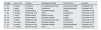
Ten CT-peritoneography evaluations performed on 10 patients with PD between January 2015 and December 2024 were reviewed (Table 1). Six patients were male, and 4 were female, with a mean age of 62.5 years (44–80) and a mean duration on PD of 5.2 months (1 week–18 months).
Patient characteristics.
| Sex/age | Time on PD | Problem | Radiological finding | Treatment (tt) | Evolution |
|---|---|---|---|---|---|
| F - 48 | 5 months | UF Failure | No vanishing point seen | Conservative intent | Changed to HD |
| M - 80 | 1 week | UF Failure | Hydrothorax | Rejected treatment | Changed to HD |
| M - 44 | 4 months | Genital edema | Dialysate leakage | Surgery | Continued on PD |
| F - 63 | 2 months | Abdominal lump | Hernia | Conservative | Continued on PD |
| M - 77 | 1 month | Genital edema | Bilateral hernias | Surgery | Continued on PD |
| M - 75 | 2 months | Genital edema | Dialysate leakage | Surgery | Continued on PD |
| M - 67 | 1 month | Abdominal lump | No vanishing point seen | Conservative | Continued on PD |
| M - 53 | 18 months | Genital edema | Dialysate leakage | Conservative | Continued on PD |
| F - 58 | 6 months | Abdominal lump | Dialysate leakage | Rejected treatment | Changed to HD |
| F - 61 | 11 months | Abdominal lump | Hernia | Conservative | Continued on PD |
PD: peritoneal dialysis; UF: ultrafiltration; HD: hemodialysis.
CT-peritoneography was performed mostly to rule out hernias and dialysate leaks. Clinically, the patients presented with 4 genital edemas, 4 abdominal lumps and 2 ultrafiltration defects.
The average time of ambulation from the infusion of the dialysate with contrast until the performance of the CT-peritoneography was 30 min, and the duration of the radiological test was less than 5 min.
Two of the abdominal lumps were identified as hernias (Fig. 1A), another was identified as dialysate leakage through the catheter entry point (Fig. 1B), and in the fourth case, the leakage point was not visualized. Cases of genital edema were identified as dialysate leakage on 3 occasions (Fig. 1C and D) and as inguinal hernias on the fourth (Fig. 1E). In one of the ultrafiltration defect cases, a dialysate leak point could not be confirmed, and in the other, a pleuroperitoneal communication was demonstrated (Fig. 1F).
. B: Leakage of dialysate through the subcutaneous route. C: Dialysate leakage due to persistence of the duct processus vaginalis. D: Leakage of the dialysate by dissection of planes from a small point of leakage (arrow). E: Genital edema due to bilateral hernias (arrows). F: Ultrafiltration failure due to peritoneal pleural leakage (arrow = vanishing point) (star = pleural effusion).")
A: Abdominal bulge due to hernia (arrow). B: Leakage of dialysate through the subcutaneous route. C: Dialysate leakage due to persistence of the duct processus vaginalis. D: Leakage of the dialysate by dissection of planes from a small point of leakage (arrow). E: Genital edema due to bilateral hernias (arrows). F: Ultrafiltration failure due to peritoneal pleural leakage (arrow = vanishing point) (star = pleural effusion).
No patient experienced immediate adverse reactions after contrast infusion. Changes in the behavior of peritoneal transport were not observed in any patient.
Five patients were treated conservatively (volume reduction in exchanges with/without change to automated dialysis), and 3 patients received restorative surgical treatment. These 8 patients were able to continue with PD. The remaining 2 patients, both of whom had ultrafiltration failure, were transferred to hemodialysis as a result of their own decision.
DiscussionNoninfectious complications are relatively common in PD patients. Among these complications, hernias and dialysate leaks occur in 4%–25% of patients, and they are promoted by increased intraperitoneal pressure.1–4
The most frequent hernial points are the umbilical canal, inguinal canal, and previous surgical incisions. Dialysate leaks can occur at any point where the integrity of the peritoneal wall has been lost and are more frequent in the inguinal canal because of the persistence of the vaginalis processus, at the point of entry of the catheter in the peritoneal cavity, and in the anterior face of the abdomen.
Sometimes it is difficult to diagnose and locate the dialysate leak, which may not be evident, although it should be suspected because of a reduction in the usual ultrafiltration.6 However, the differential diagnosis of genital edema is not easy, since it may be due to infiltration of fascial planes by dialysate leakage or to the persistence of the processus vaginalis.7
Diagnosis and precise localization are crucial for the therapeutic management of patients. However, the leakage points can be very small and not noticeable on conventional CT or peritoneal scintigraphy.
The combination of radiological contrast with dialysate solution has been used for diagnostic purposes since the 1980s.7–9 Unlike the contrast agents used in the past, the contrast agents most commonly used at present for CT-peritoneography are hypo- or iso-osmolar and nonionic and have concentrations of 270–300 mg/ml.5 The mixture of these contrast agents with the dialysis solution is stable,10 and this route of administration has not been shown to cause changes in the dialytic function of the peritoneum.11
The contrast agents used in our case were those provided at each time by the radiology service. They differed in their osmolarity and amount of iodine (higher in iohexol), but we did not observe that any was superior in terms of the quality of the images obtained.
It is preferred to use dialysate with a concentration of 1.5% glucose and the lowest ultrafiltration power so as not to increase the nephrotoxicity of the contrast agent. The dosages consist of 1 ml/kg of contrast for every 30 ml/kg of dialysate,8,12 although the addition of a fixed volume of 100 cm3 of contrast agent to a bag of 2 L of dialysate simplifies the procedure.5,10 The dilution of the contrast agent in the dialysate and the short exposure (because it is drained after performing the radiological test) significantly reduce nephrotoxicity compared with the administration of intravenous contrast agents.
CT-peritoneography offers a better diagnostic yield than other alternative imaging techniques, such as conventional CT, magnetic resonance or peritoneal scintigraphy. Scintigraphy is not available in many centers, its image capture lasts several hours, and although it can be used to confirm diagnoses, it does not allow anatomical refinement of the point of leakage or hernia.12,13 Magnetic resonance with intraperitoneal contrast offers a resolution similar to that of CT-peritoneography, without receiving radiation, but this technique has limitations in terms of accessibility (it is not available in many centers and is contraindicated in patients with metallic devices), the duration of the procedure and its economic cost.14,15
In contrast, CT-peritoneography consumes little time and does not require the patient to be in a fasted state, the risk of nephrotoxicity is reduced, and the results are immediate. In addition, current radiological CT techniques, which are highly sensitive and allow three-dimensional reconstructions, facilitate therapeutic decisions by providing great anatomical detail, which is necessary, especially if a surgical solution is selected.
The limitations of our study are the small sample size and its retrospective nature. Future studies with larger samples could compare the sensitivity of different imaging techniques for the diagnosis of hernias or dialysate leaks.
In our experience, CT-peritoneography is the most useful diagnostic technique for the clinical suspicion of hernias or dialysate leaks.
The authors declare that they have no conflicts of interest.


