array:21 [
  "pii" => "X2013251410033796"
  "issn" => "20132514"
  "doi" => "10.3265/Nefrologia.pre2010.Jan.10211"
  "estado" => "S300"
  "fechaPublicacion" => "2010-01-01"
  "documento" => "article"
  "licencia" => "http://www.elsevier.com/open-access/userlicense/1.0/"
  "subdocumento" => "fla"
  "cita" => "Nefrologia (English Version). 2010;30:87-94"
  "abierto" => array:3 [
    "ES" => true
    "ES2" => true
    "LATM" => true
  ]
  "gratuito" => true
  "lecturas" => array:2 [
    "total" => 6277
    "formatos" => array:3 [
      "EPUB" => 293
      "HTML" => 5342
      "PDF" => 642
    ]
  ]
  "Traduccion" => array:1 [
    "es" => array:17 [
      "pii" => "X0211699510033799"
      "issn" => "02116995"
      "doi" => "10.3265/Nefrologia.pre2010.Jan.10211"
      "estado" => "S300"
      "fechaPublicacion" => "2010-01-01"
      "documento" => "article"
      "licencia" => "http://www.elsevier.com/open-access/userlicense/1.0/"
      "subdocumento" => "fla"
      "cita" => "Nefrologia. 2010;30:87-94"
      "abierto" => array:3 [
        "ES" => true
        "ES2" => true
        "LATM" => true
      ]
      "gratuito" => true
      "lecturas" => array:2 [
        "total" => 10061
        "formatos" => array:3 [
          "EPUB" => 278
          "HTML" => 9126
          "PDF" => 657
        ]
      ]
      "es" => array:12 [
        "idiomaDefecto" => true
        "titulo" => "Análisis clínico de una población con poliquistosis renal autosómica dominante"
        "tienePdf" => "es"
        "tieneTextoCompleto" => "es"
        "tieneResumen" => array:2 [
          0 => "es"
          1 => "en"
        ]
        "paginas" => array:1 [
          0 => array:2 [
            "paginaInicial" => "87"
            "paginaFinal" => "94"
          ]
        ]
        "titulosAlternativos" => array:1 [
          "en" => array:1 [
            "titulo" => "Clinical analysis of a population with autosomal dominant polycystic kidney disease"
          ]
        ]
        "contieneResumen" => array:2 [
          "es" => true
          "en" => true
        ]
        "contieneTextoCompleto" => array:1 [
          "es" => true
        ]
        "contienePdf" => array:1 [
          "es" => true
        ]
        "resumenGrafico" => array:2 [
          "original" => 0
          "multimedia" => array:8 [
            "identificador" => "fig1"
            "etiqueta" => "Tab.  1"
            "tipo" => "MULTIMEDIAFIGURA"
            "mostrarFloat" => true
            "mostrarDisplay" => false
            "copyright" => "Elsevier España"
            "figura" => array:1 [
              0 => array:4 [
                "imagen" => "10211108_10211_t1.jpg"
                "Alto" => 725
                "Ancho" => 1715
                "Tamanyo" => 231083
              ]
            ]
            "descripcion" => array:1 [
              "es" => "Prevalencia, edad de aparición y tiempo transcurrido desde el diagnóstico de la enfermedad hasta la aparición de las diferentes manifestaciones clínicas renales por sexo"
            ]
          ]
        ]
        "autores" => array:1 [
          0 => array:2 [
            "autoresLista" => "Pilar Fraile Gómez, P. García-Cosmes, L. Corbacho Becerra, J.M. Tabernero Romo"
            "autores" => array:4 [
              0 => array:2 [
                "nombre" => "Pilar"
                "apellidos" => "Fraile Gómez"
              ]
              1 => array:2 [
                "Iniciales" => "P."
                "apellidos" => "García-Cosmes"
              ]
              2 => array:2 [
                "Iniciales" => "L."
                "apellidos" => "Corbacho Becerra"
              ]
              3 => array:2 [
                "Iniciales" => "J.M."
                "apellidos" => "Tabernero Romo"
              ]
            ]
          ]
        ]
      ]
      "idiomaDefecto" => "es"
      "Traduccion" => array:1 [
        "en" => array:9 [
          "pii" => "X2013251410033796"
          "doi" => "10.3265/Nefrologia.pre2010.Jan.10211"
          "estado" => "S300"
          "subdocumento" => ""
          "abierto" => array:3 [
            "ES" => true
            "ES2" => true
            "LATM" => true
          ]
          "gratuito" => true
          "lecturas" => array:1 [
            "total" => 0
          ]
          "idiomaDefecto" => "en"
          "EPUB" => "https://multimedia.elsevier.es/PublicationsMultimediaV1/item/epub/X2013251410033796?idApp=UINPBA000064"
        ]
      ]
      "EPUB" => "https://multimedia.elsevier.es/PublicationsMultimediaV1/item/epub/X0211699510033799?idApp=UINPBA000064"
      "url" => "/02116995/0000003000000001/v0_201502091336/X0211699510033799/v0_201502091337/es/main.assets"
    ]
  ]
  "itemSiguiente" => array:17 [
    "pii" => "X2013251410033800"
    "issn" => "20132514"
    "doi" => "10.3265/Nefrologia.pre2009.Dic.5646"
    "estado" => "S300"
    "fechaPublicacion" => "2010-01-01"
    "documento" => "article"
    "licencia" => "http://www.elsevier.com/open-access/userlicense/1.0/"
    "subdocumento" => "fla"
    "cita" => "Nefrologia (English Version). 2010;30:95-102"
    "abierto" => array:3 [
      "ES" => true
      "ES2" => true
      "LATM" => true
    ]
    "gratuito" => true
    "lecturas" => array:2 [
      "total" => 4701
      "formatos" => array:3 [
        "EPUB" => 275
        "HTML" => 3838
        "PDF" => 588
      ]
    ]
    "en" => array:12 [
      "idiomaDefecto" => true
      "titulo" => "Correlates of potassium transport during peritoneal equilibration tests using different dialysate glucose concentrations"
      "tienePdf" => "en"
      "tieneTextoCompleto" => "en"
      "tieneResumen" => array:2 [
        0 => "en"
        1 => "es"
      ]
      "paginas" => array:1 [
        0 => array:2 [
          "paginaInicial" => "95"
          "paginaFinal" => "102"
        ]
      ]
      "titulosAlternativos" => array:1 [
        "es" => array:1 [
          "titulo" => "Análisis del transporte peritoneal de potasio mediante pruebas de equilibrio peritoneal con diferentes concentraciones de glucosa"
        ]
      ]
      "contieneResumen" => array:2 [
        "en" => true
        "es" => true
      ]
      "contieneTextoCompleto" => array:1 [
        "en" => true
      ]
      "contienePdf" => array:1 [
        "en" => true
      ]
      "resumenGrafico" => array:2 [
        "original" => 0
        "multimedia" => array:8 [
          "identificador" => "fig1"
          "etiqueta" => "Tab.  1"
          "tipo" => "MULTIMEDIAFIGURA"
          "mostrarFloat" => true
          "mostrarDisplay" => false
          "copyright" => "Elsevier España"
          "figura" => array:1 [
            0 => array:4 [
              "imagen" => "1003618078_t1_pag97.jpg"
              "Alto" => 382
              "Ancho" => 403
              "Tamanyo" => 43782
            ]
          ]
          "descripcion" => array:1 [
            "en" => "Study population"
          ]
        ]
      ]
      "autores" => array:1 [
        0 => array:2 [
          "autoresLista" => "A.M. Gomes, A. Rodríguez-Carmona, Miguel Pérez Fontán, A. López Muñiz, N. Blanco Castro, M. da Cunha, T. García Falcón"
          "autores" => array:7 [
            0 => array:2 [
              "Iniciales" => "A.M."
              "apellidos" => "Gomes"
            ]
            1 => array:2 [
              "Iniciales" => "A."
              "apellidos" => "Rodríguez-Carmona"
            ]
            2 => array:2 [
              "nombre" => "Miguel"
              "apellidos" => "Pérez Fontán"
            ]
            3 => array:2 [
              "Iniciales" => "A."
              "apellidos" => "López Muñiz"
            ]
            4 => array:2 [
              "Iniciales" => "N."
              "apellidos" => "Blanco Castro"
            ]
            5 => array:2 [
              "Iniciales" => "M."
              "apellidos" => "da Cunha"
            ]
            6 => array:2 [
              "Iniciales" => "T."
              "apellidos" => "García Falcón"
            ]
          ]
        ]
      ]
    ]
    "idiomaDefecto" => "en"
    "Traduccion" => array:1 [
      "es" => array:9 [
        "pii" => "X0211699510033803"
        "doi" => "10.3265/Nefrologia.pre2009.Dic.5646"
        "estado" => "S300"
        "subdocumento" => ""
        "abierto" => array:3 [
          "ES" => true
          "ES2" => true
          "LATM" => true
        ]
        "gratuito" => true
        "lecturas" => array:1 [
          "total" => 0
        ]
        "idiomaDefecto" => "es"
        "EPUB" => "https://multimedia.elsevier.es/PublicationsMultimediaV1/item/epub/X0211699510033803?idApp=UINPBA000064"
      ]
    ]
    "EPUB" => "https://multimedia.elsevier.es/PublicationsMultimediaV1/item/epub/X2013251410033800?idApp=UINPBA000064"
    "url" => "/20132514/0000003000000001/v0_201502091608/X2013251410033800/v0_201502091609/en/main.assets"
  ]
  "itemAnterior" => array:17 [
    "pii" => "X2013251410033788"
    "issn" => "20132514"
    "doi" => "10.3265/Nefrologia.pre2009.Dic.5732"
    "estado" => "S300"
    "fechaPublicacion" => "2010-01-01"
    "documento" => "article"
    "licencia" => "http://www.elsevier.com/open-access/userlicense/1.0/"
    "subdocumento" => "fla"
    "cita" => "Nefrologia (English Version). 2010;30:78-86"
    "abierto" => array:3 [
      "ES" => true
      "ES2" => true
      "LATM" => true
    ]
    "gratuito" => true
    "lecturas" => array:2 [
      "total" => 9282
      "formatos" => array:3 [
        "EPUB" => 310
        "HTML" => 6649
        "PDF" => 2323
      ]
    ]
    "en" => array:11 [
      "idiomaDefecto" => true
      "titulo" => "Prevalence of chronic renal disease in Spain: Results of the EPIRCE study"
      "tienePdf" => "en"
      "tieneTextoCompleto" => "en"
      "tieneResumen" => array:2 [
        0 => "en"
        1 => "es"
      ]
      "paginas" => array:1 [
        0 => array:2 [
          "paginaInicial" => "78"
          "paginaFinal" => "86"
        ]
      ]
      "contieneResumen" => array:2 [
        "en" => true
        "es" => true
      ]
      "contieneTextoCompleto" => array:1 [
        "en" => true
      ]
      "contienePdf" => array:1 [
        "en" => true
      ]
      "resumenGrafico" => array:2 [
        "original" => 0
        "multimedia" => array:8 [
          "identificador" => "fig1"
          "etiqueta" => "Tab.  2"
          "tipo" => "MULTIMEDIAFIGURA"
          "mostrarFloat" => true
          "mostrarDisplay" => false
          "copyright" => "Elsevier España"
          "figura" => array:1 [
            0 => array:4 [
              "imagen" => "1011718078_t2_pag83.jpg"
              "Alto" => 567
              "Ancho" => 829
              "Tamanyo" => 92459
            ]
          ]
          "descripcion" => array:1 [
            "en" => "Prevalence of chronic kidney disease in the Spanish population aged 20 years or older based on the cohort collected in the EPIRCE study (n = 2,746)"
          ]
        ]
      ]
      "autores" => array:1 [
        0 => array:2 [
          "autoresLista" => " on behalf of the EPIRCE Study Group, Alfonso Otero González, A. de Francisco, P. Gayoso, F. García"
          "autores" => array:5 [
            0 => array:1 [
              "apellidos" => "on behalf of the EPIRCE Study Group"
            ]
            1 => array:2 [
              "nombre" => "Alfonso"
              "apellidos" => "Otero González"
            ]
            2 => array:2 [
              "Iniciales" => "A."
              "apellidos" => "de Francisco"
            ]
            3 => array:2 [
              "Iniciales" => "P."
              "apellidos" => "Gayoso"
            ]
            4 => array:2 [
              "Iniciales" => "F."
              "apellidos" => "García"
            ]
          ]
        ]
      ]
    ]
    "idiomaDefecto" => "en"
    "Traduccion" => array:1 [
      "en" => array:9 [
        "pii" => "X0211699510033780"
        "doi" => "10.3265/Nefrologia.pre2009.Dic.5732"
        "estado" => "S300"
        "subdocumento" => ""
        "abierto" => array:3 [
          "ES" => true
          "ES2" => true
          "LATM" => true
        ]
        "gratuito" => true
        "lecturas" => array:1 [
          "total" => 0
        ]
        "idiomaDefecto" => "en"
        "EPUB" => "https://multimedia.elsevier.es/PublicationsMultimediaV1/item/epub/X0211699510033780?idApp=UINPBA000064"
      ]
    ]
    "EPUB" => "https://multimedia.elsevier.es/PublicationsMultimediaV1/item/epub/X2013251410033788?idApp=UINPBA000064"
    "url" => "/20132514/0000003000000001/v0_201502091608/X2013251410033788/v0_201502091609/en/main.assets"
  ]
  "en" => array:15 [
    "idiomaDefecto" => true
    "titulo" => "Clinical analysis of a population with autosomal dominant polycystic kidney disease"
    "tieneTextoCompleto" => true
    "paginas" => array:1 [
      0 => array:2 [
        "paginaInicial" => "87"
        "paginaFinal" => "94"
      ]
    ]
    "autores" => array:1 [
      0 => array:3 [
        "autoresLista" => "Pilar Fraile Gómez, P. García-Cosmes, L. Corbacho Becerra, J.M. Tabernero Romo"
        "autores" => array:4 [
          0 => array:4 [
            "nombre" => "Pilar"
            "apellidos" => "Fraile Gómez"
            "email" => array:1 [
              0 => "pilarfg@usal.es"
            ]
            "referencia" => array:1 [
              0 => array:2 [
                "etiqueta" => "<span class="elsevierStyleSup">a</span>"
                "identificador" => "affa"
              ]
            ]
          ]
          1 => array:3 [
            "Iniciales" => "P."
            "apellidos" => "Garc&#237;a-Cosmes"
            "referencia" => array:1 [
              0 => array:2 [
                "etiqueta" => "<span class="elsevierStyleSup">a</span>"
                "identificador" => "affa"
              ]
            ]
          ]
          2 => array:3 [
            "Iniciales" => "L."
            "apellidos" => "Corbacho Becerra"
            "referencia" => array:1 [
              0 => array:2 [
                "etiqueta" => "<span class="elsevierStyleSup">a</span>"
                "identificador" => "affa"
              ]
            ]
          ]
          3 => array:3 [
            "Iniciales" => "J.M."
            "apellidos" => "Tabernero Romo"
            "referencia" => array:1 [
              0 => array:2 [
                "etiqueta" => "<span class="elsevierStyleSup">a</span>"
                "identificador" => "affa"
              ]
            ]
          ]
        ]
        "afiliaciones" => array:1 [
          0 => array:3 [
            "entidad" => "Servicio de Nefrología, Hospital Universitario de Salamanca, Salamanca,  España, "
            "etiqueta" => "<span class="elsevierStyleSup">a</span>"
            "identificador" => "affa"
          ]
        ]
      ]
    ]
    "titulosAlternativos" => array:1 [
      "es" => array:1 [
        "titulo" => "An&#225;lisis cl&#237;nico de una poblaci&#243;n con poliquistosis renal autos&#243;mica dominante"
      ]
    ]
    "resumenGrafico" => array:2 [
      "original" => 0
      "multimedia" => array:7 [
        "identificador" => "fig1"
        "etiqueta" => "Fig. 1"
        "tipo" => "MULTIMEDIAFIGURA"
        "mostrarFloat" => true
        "mostrarDisplay" => false
        "copyright" => "Elsevier Espa&#241;a"
        "figura" => array:1 [
          0 => array:4 [
            "imagen" => "1021118078_f1_pag89.jpg"
            "Alto" => 425
            "Ancho" => 826
            "Tamanyo" => 30937
          ]
        ]
      ]
    ]
    "textoCompleto" => "<p class="elsevierStylePara"><span class="elsevierStyleBold">INTRODUCTION</span></p><p class="elsevierStylePara">Autosomal dominant polycystic kidney disease &#40;ADPKD&#41; is a hereditary multiorgan disease characterised by the progressive growth and development of renal cysts that destroy functional parenchyma&#46; It affects one in every 1&#44;000 people and is responsible for the 7-10&#37; of cases of chronic kidney disease which require renal replacement treatment&#46;<span class="elsevierStyleSup">1</span> ADPKD is caused by a mutation in the <span class="elsevierStyleItalic">PKD1</span> gene&#44; responsible for 85-90&#37; of cases&#44; in the <span class="elsevierStyleItalic">PKD2</span> gene&#44; responsible for 10-15&#37; of cases and&#44; possibly in a third gene&#44; <span class="elsevierStyleItalic">PKD3</span>&#44; which has not yet been identified&#46;<span class="elsevierStyleSup">2-4</span></p><p class="elsevierStylePara">ADPKD is a multisystemic disease&#44; with renal and extrarenal manifestations derived from the formation of renal cysts and&#44; in many cases&#44; asymptomatic cysts in the liver and pancreas&#46;<span class="elsevierStyleSup">5</span> The main renal symptoms are abdominal or lumbar pain&#44; haematuria&#44; arterial hypertension &#40;AH&#41; and&#44; less frequently&#44; urinary infection&#44; lithiasis&#44; renal failure and palpable renal mass&#46;<span class="elsevierStyleSup">6</span> The main extrarenal manifestations are cerebral aneurysms&#44; hepatic cysts&#44; cardiac valve disease&#44; colonic diverticula and abdominal hernias&#46;<span class="elsevierStyleSup">7&#44;8</span></p><p class="elsevierStylePara">Diagnosis of ADPKD is usually established by means of imaging techniques&#59; renal echography is the method of choice in patients over 30 years of age&#46; In younger patients computed tomography &#40;CAT&#41; or nuclear magnetic resonance &#40;NMR&#41; should be used&#46;<span class="elsevierStyleSup">9</span> In patients under 30&#44; diagnostic certainty is offered by genetic study which provides additional information in genetic counseling&#46;<span class="elsevierStyleSup">10&#44;11</span></p><p class="elsevierStylePara">In this study&#44; our purpose was to analyse the clinical characteristics of all ADPKD patients who required medical attention in our centre during the 1994-2005 period&#46;</p><p class="elsevierStylePara"><span class="elsevierStyleBold">MATERIAL AND METHODS</span></p><p class="elsevierStylePara"><span class="elsevierStyleBold">Study population</span></p><p class="elsevierStylePara">We carried out a clinical study of 48 ADPKD patients who required medical attention in the Nephrology Service of the University Hospital of Salamanca between 1994 and 2005&#46; The patients had been diagnosed with ADPKD according to clinical and radiological criteria&#46;</p><p class="elsevierStylePara"><span class="elsevierStyleBold">Clinical follow-up</span></p><p class="elsevierStylePara">The clinical aspects of 48 patients diagnosed with ADPKD who required medical intervention were analysed by our service through the eleven years from 1994 to 2005&#44; and they were followed-up clinically until March 2008&#46; The information on all the patients was taken from their clinical records&#46; The principal parameters analysed were prevalence and age of onset of the disease&#44; aspects related to its initial clinical manifestations&#44; to the clinical manifestations that appeared during evolution&#44; to extrarenal manifestations and to the survival and morbidity and mortality of patients diagnosed with ADPKD&#46;</p><p class="elsevierStylePara"><span class="elsevierStyleBold">Statistical method</span></p><p class="elsevierStylePara">The statistical study was conducted with the SPSS<span class="elsevierStyleSup">&#174;</span> software package for Mac<span class="elsevierStyleSup">&#174;</span> OS X &#40;version 13&#46;0&#41;&#46; Qualitative variables were expressed as percentages and numeric variables as mean and standard deviation &#40;X &#177; SD&#41;&#46;</p><p class="elsevierStylePara">The statistical method used was the Student&#8217;s t-test to compare hypothesis on means in populations with normal distribution&#46; When the principle of normality was not met the Mann-Whitney non-parametric statistical test was used&#46; The relation between the qualitative variables of two or more categories was determined by proportion using the Chi-squared test&#46;</p><p class="elsevierStylePara">Independently of the analysis and the nature of the variable&#44; probability values of less than 0&#46;05 &#40;p &#60; 0&#46;05&#41; were considered to be significant&#46;</p><p class="elsevierStylePara"><span class="elsevierStyleBold">RESULTS</span></p><p class="elsevierStylePara">48 ADPKD patients&#44; 21 women and 27 men&#44; were clinically studied&#46; The average age of onset of the first symptoms was 46&#46;07 &#177; 13&#46;49 years&#44; 41&#46;17 &#177; 13&#46;41 years in women and 49&#46;91 &#177; 12&#46;52 years in men &#40;p &#60; 0&#46;05&#41;&#46; 41&#46;18&#37; of the women had the first manifestations of the disease between 30 and 40 years of age while diagnosis in men was fundamentally between 50 and 60 years&#46;</p><p class="elsevierStylePara">No statistically significant differences were found in the prevalence of the disease according to the sex of the patient&#46;</p><p class="elsevierStylePara"><span class="elsevierStyleBold">Initial clinical renal manifestations</span></p><p class="elsevierStylePara">As an isolated manifestation&#44; AH was the most frequent disorder and the initial sign of the disease in 68&#46;42&#37; of the patients&#44; predominantly in men with respect to women &#40;p &#60; 0&#46;05&#41;&#44; followed by chronic renal failure &#40;CRF&#41;&#44; present in 68&#46;29&#37; of patients with ADPKD &#40;Figure 1&#41;&#46;</p><p class="elsevierStylePara">No statistically significant differences were found in the prevalence of the different initial clinical renal manifestations by gender&#44; with the exception of AH&#44; proteinuria&#44; hyperuricaemia and renal lithiasis in which the prevalence was greater in men &#40;p &#60; 0&#46;05&#41; than in women&#46; Quantification of proteinuria at the time of diagnosis was always less than 300mg&#47;24 h&#46;</p><p class="elsevierStylePara"><span class="elsevierStyleBold">Clinical renal manifestations during evolution </span></p><p class="elsevierStylePara">The most frequent renal manifestation during evolution was CRF&#44; present in 100&#37; of the studied patients&#44; followed by proteinuria &#40;92&#46;31&#37;&#41; and terminal chronic renal failure &#40;TCRF&#41; &#40;89&#46;58 &#37;&#41; &#40;Table 1&#41;&#46; AH was present in 87&#46;23&#37; of the sick patients&#44; statistically significant in the 30-60 age group &#40;p &#60; 0&#46;01&#41;&#46; The prevalence of renal manifestations during evolution showed no significant differences between men and women &#40;Table 1&#41;&#46;</p><p class="elsevierStylePara">Although the prevalence of urinary tract infections was higher in women&#44; it had no statistical significance&#46; Enterobacteria were responsible for 94&#46;12&#37; of the episodes and <span class="elsevierStyleItalic">Escherichia coli </span>was the most frequently isolated aetiological agent &#40;70&#46;59&#37;&#41;&#46;</p><p class="elsevierStylePara">The mean age at CRF onset was 50&#46;02 &#177; 11&#46;21 years&#44; and that of TCRF 54&#46;74 &#177; 10&#46;07 years&#44; independently of sex &#40;p &#62; 0&#46;05&#41; &#40;Table 1&#41;&#46; Patients diagnosed with CRF were found predominantly in the 30-60 age group &#40;p &#60; 0&#46;05&#41;&#44; as were patients with TCRF&#44; in whom the difference was not statistically significant&#46; When the disease was transmitted by the father&#44; the mean age of onset of CRF and of TCRF was lower &#40;p &#60; 0&#46;01&#41;&#46;</p><p class="elsevierStylePara">The evolution time to CRF and TCRF &#40;Figure 2&#41; from diagnosis of the disease was shorter in men than in women&#44; as was the elapsed time from diagnosis of CRF to the start of renal replacement therapy &#40;p &#60; 0&#46;05&#41; &#40;Table 1 and Figure 3&#41;&#46; Patients with AH were more likely to exhibit CRF &#40;p &#60; 0&#46;05&#41;&#46;</p><p class="elsevierStylePara">Of all the patients who started renal replacement therapy&#44; 83&#46;72&#37; were hypertensive and 13&#46;95&#37; normotensive&#46; Patients with AH&#44; proteinuria&#44; macroscopic haematuria&#44; lithiasis&#44; abdominal pain&#44; urinary tract infections&#44; hepatic cysts and cyst infections were more likely to exhibit TCRF &#40;p &#60; 0&#46;01&#41;&#46; However&#44; the probability of developing TCRF did not increase in patients with diverticula&#44; macroscopic haematuria before 30 years of age or women with more than 3 children &#40;p &#62; 0&#46;05&#41;&#46;</p><p class="elsevierStylePara">No significant differences were found by sex in the age of onset of any of the renal manifestations &#40;Table 1&#41;&#46;</p><p class="elsevierStylePara">In addition to CRF and TCRF&#44; the time of evolution from disease diagnosis to the appearance of hyperuricaemia&#44; proteinuria&#44; renal lithiasis and AH was also less in men than in women &#40;p &#60; 0&#46;05&#41; &#40;Table 1 and Figure 3&#41;&#46; No significant differences were found in the evolution period from diagnosis of the disease to the appearance of the other renal manifestations&#46;</p><p class="elsevierStylePara">The recurrence of renal lithiasis was statistically significant in men&#46;</p><p class="elsevierStylePara"><span class="elsevierStyleBold">Extrarenal manifestations</span></p><p class="elsevierStylePara">Hepatic cysts were the most frequent extrarenal manifestation&#44; present at the time of diagnosis in 59&#46;09&#37; of patients&#44; with greater prevalence in women &#40;p &#60; 0&#46;05&#41; &#40;Figure 4&#41;&#46; No statistically significant differences by sex were found in the prevalence of the other extrarenal manifestations diagnosed&#46;</p><p class="elsevierStylePara"><span class="elsevierStyleBold">Survival and morbidity and mortality</span></p><p class="elsevierStylePara">Patients with ADPKD had a total of 6&#46;17 &#177; 5&#46;81 hospital admissions in our centre during the length of the study&#44; 0&#46;94 &#177; 0&#46;63 admissions&#47;year&#44; with an average stay of 11&#46;27 &#177; 4&#46;91 days&#46; No significant differences by sex were found in survival&#44; or in the number of hospital admissions per year&#44; nor in the average stay &#40;Table 2&#41;&#46;</p><p class="elsevierStylePara">Of the total number of patients with ADPKD&#44; 39&#46;58&#37; died during the study&#44; 29&#46;17&#37; men and 10&#46;42&#37; women &#40;p &#60; 0&#46;05&#59; RR &#61; 2&#46;3&#41;&#46; Patient survival from the time of diagnosis was 13&#46;4 &#177; 7&#46;79 years&#44; but it was not shown that AH&#44; macroscopic haematuria&#44; abdominal pain&#44; cyst infections&#44; renal lithiasis&#44; urinary infections&#44; the presence of hepatic cysts or a large number of childbirths were risk factors for mortality &#40;p &#62; 0&#46;05&#41;&#46;</p><p class="elsevierStylePara">The principal causes of death were of cardiovascular &#40;42&#46;1&#37;&#41; and infectious &#40;42&#46;1&#37;&#41; origin&#46;</p><p class="elsevierStylePara"><span class="elsevierStyleBold">DISCUSSION</span></p><p class="elsevierStylePara"><span class="elsevierStyleBold">CLINICAL DIAGNOSIS</span></p><p class="elsevierStylePara">ADPKD is diagnosed in the majority of patients by characteristic clinical manifestations which usually appear in the third or fourth decade of life&#46; In our study&#44; the mean age of onset of the first clinical manifestations of ADPKD was earlier in women than in men&#46; Studies published to date have not analysed the existence of differences by sex in age at disease diagnosis or in the prevalence of the majority of renal and extrarenal manifestations&#46;<span class="elsevierStyleSup">1&#44;4&#44;7&#44;9&#44;12</span></p><p class="elsevierStylePara">In this series&#44; the most frequent initial manifestation at diagnosis was AH&#44; present in 68&#46;42&#37; of patients&#44; with predominance in males and with a mean age of onset &#40;45&#46;77 &#177; 10&#46;48 years&#41; later than that described in the literature&#44; with a mean age of 30 years&#59;<span class="elsevierStyleSup">13&#44;14</span> nevertheless&#44; in our study it preceded measurable changes in renal function by several years&#46;<span class="elsevierStyleSup">1&#44;13&#44;15</span> AH is&#44; according to different publications&#44; one of the guiding signs for ADPKD diagnosis&#44; which is confirmed by this study&#46; The fact that AH diagnosis in our population was later than that published<span class="elsevierStyleSup">1&#44;14&#44;16</span> could be explained by the current saturation of general medical consultations and by the lack of protocolised studies which clarify AH aetiology when it is light or easy to control&#59; cases of ADPKD would thus be disguised among those in which early and vigorous treatment of complications would delay the onset of CRF and the progression of TCRF&#46; The high prevalence in our study of CRF and TCRF secondary to ADPKD is in accordance with the ways the disease presents&#46; During the evolution of ADPKD&#44; AH was present in 87&#46;23&#37; of our patients&#44; with no differences observed between the sexes&#46; Evolution time from disease diagnosis to the appearance of AH was less in men&#46; Its role in the progression of renal failure&#44;<span class="elsevierStyleSup">13&#44;17</span> already described&#44; was demonstrated&#59; we were unable to confirm this in patients under 35 given the low prevalence of young patients in our study&#46; It has been proved&#44; both in the general population and in sufferers of ADPKD that the prevalence of AH is significantly greater in men up to 45 years of age&#44; at which point&#44; although still prevalent in men&#44; the differences are no longer significant&#46; The shorter evolution time from disease diagnosis to AH onset in men may be due to the existence of cardiovascular risk factors &#40;smoking habit&#44; dyslipidaemia&#44; central obesity&#41; that favour the development of AH and that predominate in males between the ages of 20 and 55&#46; From there on&#44; hormonal influences&#44; dyslipidaemia or central obesity cause these factors to increase significantly in women and&#44; as a result&#44; so does AH&#46;<span class="elsevierStyleSup">14</span></p><p class="elsevierStylePara">In this series&#44; proteinuria was one of the most frequent renal manifestations of ADPKD at the time of diagnosis&#44; following AH&#44; renal failure and haematuria&#46; It was the presenting sign of the disease in 36&#37; of the men&#44; statistically significant with respect to the women&#44; and the elapsed time from disease diagnosis to onset of proteinuria was shorter in men&#46; AH and proteinuria present at the time of diagnosis has been related to worse renal prognosis and increased cardiovascular risk in ADPKD&#46;<span class="elsevierStyleSup">17&#44;18</span> Given that AH is correlated to the onset of proteinuria in various publications&#44;<span class="elsevierStyleSup">19</span> the greater prevalence of AH in men at the time of diagnosis in this study would also explain the greater prevalence of proteinuria&#46; Furthermore&#44; AH&#44; dyslipidaemia&#44; central obesity and tobacco habit increase the risk of proteinuria and predominate in middle-aged men&#46; During disease evolution&#44; proteinuria becomes the second most frequent renal manifestation &#40;92&#46;31&#37;&#41; with no gender difference&#44; which would explain the high percentage of patients with established kidney damage&#46; The quantification of proteinuria was always lower than 300mg&#47;dL&#44; which agrees with that already described<span class="elsevierStyleSup">19</span> and there were no cases of nephrotic-range proteinuria&#46; In the ADPKD patients with proteinuria studied&#44; it was shown that they were more likely to present TCRF&#44; in relation to secondary glomerular hyperfiltration&#46;</p><p class="elsevierStylePara">Haematuria was a symptom presenting the disease in 47&#46;06&#37; of the cases&#44; and was present in the evolution of the disease in 88&#46;89&#37; of our ADPKD patients&#46; This prevalence was superior to that reported in literature&#44; where the figure is 35-50&#37;&#46;<span class="elsevierStyleSup">1</span> Haematuria could be due to renal calculus&#44; cyst infection or malignant tumours&#46; The elevated prevalence of renal lithiasis and cyst infections in our series explains the higher prevalence of haematuria than that described&#46; The underlying association of macroscopic haematuria with the progression of renal failure<span class="elsevierStyleSup">17&#44;20</span> was evident&#44; but that role was not demonstrated in patients under 30 probably due to the low prevalence of young patients with ADPKD&#46; This deleterious effect of haematuria on kidney function can be explained both by the obstructive episodes secondary to clot formation and the hypovolaemic situations that it provokes&#46;</p><p class="elsevierStylePara">The prevalence of urinary tract infections in our study was similar to that described by others&#46; As in the general population&#44; because the female urethra is shorter&#44; prevalence was higher in women&#44; but was not statistically significant&#46;<span class="elsevierStyleSup">12&#44;16</span><span class="elsevierStyleItalic">E&#46; coli</span> was the most frequently isolated aetiological agent&#46; The most frequent infections were cyst infections and acute pyelonephritis&#44; with a determinant role in the progression of renal failure&#44; probably in relation to the deterioration of kidney function secondary to episodes of haemodynamic instability derived from severe infection&#46;</p><p class="elsevierStylePara">CRF was the second most frequent clinical manifestation at diagnosis and that of greatest prevalence in the evolution of the disease in our study&#44; while in other series it is estimated that only around half of the patients with mutation will develop the disease before the sixth decade of life&#46;<span class="elsevierStyleSup">15</span> Also&#44; the need for renal replacement treatment in our work was higher than that described&#46;<span class="elsevierStyleSup">18</span> The mean age of onset of CRF &#40;50&#46;02 &#177; 11&#46;21 years&#41; and TCRF &#40;54&#46;74 &#177; 10&#46;07 years&#41; was similar to that described in the literature for patients with <span class="elsevierStyleItalic">PKD1</span> gene mutation and requiring renal replacement treatment &#40;54&#46;3 years&#41;&#44;<span class="elsevierStyleSup">3&#44;4</span> which leads us to assume a greater prevalence of patients in our population with <span class="elsevierStyleItalic">PKD1</span> gene mutation if we limit ourselves to a clinical analysis&#46; The age of onset of TCRF was earlier when transmitted by the father than by the mother and evolution time to CRF and TCRF from disease diagnosis was shorter in men than in women&#44; a fact already reflected in different studies&#46;<span class="elsevierStyleSup">3&#44;21</span> In this study&#44; men had more probability of rapid progression to renal failure&#44; which appears to be related to a direct effect of the sexual hormones&#46;<span class="elsevierStyleSup">21&#44;22</span> Another sign of poor prognosis in the early evolution to both CRF and TCRF was the disease being inherited from the father&#46;</p><p class="elsevierStylePara">As far as extrarenal manifestations&#44;<span class="elsevierStyleSup">8</span> these predominated in women&#44; although only hepatic cysts were statistically significant&#58; the greater prevalence of these in women has been associated with the number of pregnancies&#44; the taking of contraceptive pills and replacement hormonal therapy&#46; Although there are contradictory results&#44; we confirm that people with hepatic cysts have worse renal function than those who do not&#44;<span class="elsevierStyleSup">3&#44;16</span> probably due to the prejudicial effect that repeated infection in hepatic cysts would have on renal function&#46;</p><p class="elsevierStylePara">In this study&#44; a similar prevalence was observed in deaths of cardiovascular &#40;42&#46;1&#37;&#41; and infectious &#40;42&#46;1&#37;&#41; origin&#46; Before 1975&#44; the most frequent cause of death in ADPKD patients was infection &#40;30&#37;&#41;&#44; uraemia &#40;28&#37;&#41; and cardiovascular disease &#40;21&#37;&#41;&#46; With the development of dialysis&#44; the main cause of mortality has become cardiovascular &#40;34&#37;&#41; followed by infections &#40;20&#46;4&#37;&#41;&#46;<span class="elsevierStyleSup">23</span> The principal cause of death of patients in renal replacement treatment and those subjected to renal transplant is cardiovascular&#59; nevertheless&#44; in our study there was a markedly high prevalence of deaths of infectious origin&#44; which could be attributed to the immunodeficiency situation derived from a population with very high prevalence of TCRF secondary to ADPKD&#46; The death rate was higher in men&#44; but no significant differences were found in survival&#46;</p><p class="elsevierStylePara">In spite of the mean age of ADPKD diagnosis in this study being lower in women&#44; evolution up to the appearance of renal lithiasis&#44; hyperuricaemia&#44; proteinuria&#44; AH and TCRF was shorter in men&#46; This&#44; together with the greater prevalence in men&#44; at diagnosis&#44; of risk factors involved in the progression towards TCRF&#44; determined a more torpid evolution of the disease in men&#44; an earlier start of renal replacement treatment and&#44; consequently&#44; greater mortality&#46; In women&#44; however&#44; the evolution time of the disease was longer&#44; which translated to a higher number of hospital admissions&#46; We may think that in women morbidity predominates and in men&#44; mortality&#44; and it is probably not a reflection of the disease but an observation common to the entire population&#46;</p><p class="elsevierStylePara">In short&#44; in our study&#44; male carriers of ADPKD were diagnosed later than women&#44; which could explain their worse clinical evolution&#46; In this population&#44; AH was the most frequent clinical manifestation at diagnosis&#44; and this classification should&#44; therefore&#44; be included in all cases with unknown AH&#46; Also&#44; infectious and cardiovascular complications were the principal cause of mortality&#44; implying that infectious complications are a sign of alert in all ADPKD patients&#44; including those not subjected to transplant&#46;</p><p class="elsevierStylePara"><a href="grande&#47;1021118078&#95;f1&#95;pag89&#46;jpg" class="elsevierStyleCrossRefs"><img src="1021118078_f1_pag89.jpg"></img></a></p><p class="elsevierStylePara">Figure 1&#46; </p><p class="elsevierStylePara"><a href="grande&#47;1021118078&#95;t1&#95;pag90&#46;jpg" class="elsevierStyleCrossRefs"><img src="1021118078_t1_pag90.jpg" alt="Prevalence&#44; age of appearance and elapsed time from disease diagnosis to the appearance of the different renal clinical manifestations by sex"></img></a></p><p class="elsevierStylePara">Table 1&#46; Prevalence&#44; age of appearance and elapsed time from disease diagnosis to the appearance of the different renal clinical manifestations by sex</p><p class="elsevierStylePara"><a href="grande&#47;1021118078&#95;f2&#95;pag90&#46;jpg" class="elsevierStyleCrossRefs"><img src="1021118078_f2_pag90.jpg"></img></a></p><p class="elsevierStylePara">Figure 2&#46; </p><p class="elsevierStylePara"><a href="grande&#47;1021118078&#95;f3&#95;pag91&#46;jpg" class="elsevierStyleCrossRefs"><img src="1021118078_f3_pag91.jpg"></img></a></p><p class="elsevierStylePara">Figure 3&#46; </p><p class="elsevierStylePara"><a href="grande&#47;1021118078&#95;t2&#95;pag91&#46;jpg" class="elsevierStyleCrossRefs"><img src="1021118078_t2_pag91.jpg" alt="Prevalence and statistical significance by sex of deceased patients&#44; mean age at time of death&#44; survival since diagnosis&#44; hospital admissions and mean stay"></img></a></p><p class="elsevierStylePara">Table 2&#46; Prevalence and statistical significance by sex of deceased patients&#44; mean age at time of death&#44; survival since diagnosis&#44; hospital admissions and mean stay</p><p class="elsevierStylePara"><a href="grande&#47;1021118078&#95;f4&#95;pag91&#46;jpg" class="elsevierStyleCrossRefs"><img src="1021118078_f4_pag91.jpg"></img></a></p><p class="elsevierStylePara">Figure 4&#46; </p>"
    "pdfFichero" => "P1-E43-S1785-A10211-EN.pdf"
    "tienePdf" => true
    "PalabrasClave" => array:2 [
      "es" => array:6 [
        0 => array:4 [
          "clase" => "keyword"
          "titulo" => "Palabras clave"
          "identificador" => "xpalclavsec437787"
          "palabras" => array:1 [
            0 => "Poliquistosis renal autos&#243;mica dominante"
          ]
        ]
        1 => array:4 [
          "clase" => "keyword"
          "titulo" => "Palabras clave"
          "identificador" => "xpalclavsec437789"
          "palabras" => array:1 [
            0 => "PKD2"
          ]
        ]
        2 => array:4 [
          "clase" => "keyword"
          "titulo" => "Palabras clave"
          "identificador" => "xpalclavsec437791"
          "palabras" => array:1 [
            0 => "Insuficiencia renal cr&#243;nica"
          ]
        ]
        3 => array:4 [
          "clase" => "keyword"
          "titulo" => "Palabras clave"
          "identificador" => "xpalclavsec437793"
          "palabras" => array:1 [
            0 => "PKD1"
          ]
        ]
        4 => array:4 [
          "clase" => "keyword"
          "titulo" => "Palabras clave"
          "identificador" => "xpalclavsec437795"
          "palabras" => array:1 [
            0 => "Insuficiencia renal cr&#243;nica terminal"
          ]
        ]
        5 => array:4 [
          "clase" => "keyword"
          "titulo" => "Palabras clave"
          "identificador" => "xpalclavsec437797"
          "palabras" => array:1 [
            0 => "Hipertensi&#243;n arterial"
          ]
        ]
      ]
      "en" => array:6 [
        0 => array:4 [
          "clase" => "keyword"
          "titulo" => "Keywords"
          "identificador" => "xpalclavsec437788"
          "palabras" => array:1 [
            0 => "Autosomal dominant polycystic kidney disease"
          ]
        ]
        1 => array:4 [
          "clase" => "keyword"
          "titulo" => "Keywords"
          "identificador" => "xpalclavsec437790"
          "palabras" => array:1 [
            0 => "PKD2"
          ]
        ]
        2 => array:4 [
          "clase" => "keyword"
          "titulo" => "Keywords"
          "identificador" => "xpalclavsec437792"
          "palabras" => array:1 [
            0 => "Chronic renal failure"
          ]
        ]
        3 => array:4 [
          "clase" => "keyword"
          "titulo" => "Keywords"
          "identificador" => "xpalclavsec437794"
          "palabras" => array:1 [
            0 => "PKD1"
          ]
        ]
        4 => array:4 [
          "clase" => "keyword"
          "titulo" => "Keywords"
          "identificador" => "xpalclavsec437796"
          "palabras" => array:1 [
            0 => "End-stage renal failure"
          ]
        ]
        5 => array:4 [
          "clase" => "keyword"
          "titulo" => "Keywords"
          "identificador" => "xpalclavsec437798"
          "palabras" => array:1 [
            0 => "Arterial hypertension"
          ]
        ]
      ]
    ]
    "tieneResumen" => true
    "resumen" => array:2 [
      "en" => array:1 [
        "resumen" => "<p class="elsevierStylePara">Autosomal dominant polycystic kidney disease is a multi-organic hereditary disorder&#46; It is responsible for 7-10&#37; of cases of end stage renal failure&#46; It is caused by mutations in the genes <span class="elsevierStyleItalic">PKD1</span> and <span class="elsevierStyleItalic">PKD2</span>&#46; The diagnosis of this disease can be performed through ultrasounds&#44; but the molecular diagnosis offers some advantages&#44; such as the early detection of asymptomatic individuals who carry this genetic defect&#44; in order to perform a preventive monitoring and genetic counselling&#46; In this work&#44; we present the results of the clinical analysis of 48 patients diagnosed with autosomal dominant polycystic kidney disease&#46; The objectives of this work were to analyze the main clinical aspects of the disease&#46; The average age of appearance of the first symptoms was 41&#46;17 &#177; 13&#46;41 years in women and 49&#46;91 &#177; 12&#46;52 years in men &#40;p &#60;0&#46;05&#41;&#46; Arterial hypertension was the first sign of the disease &#40;68&#46;42&#37;&#41;&#44; with more cases in men than in women &#40;p &#60;0&#46;05&#41;&#44; followed by chronic renal failure &#40;68&#46;29 &#37;&#41;&#46; The most common renal symptom during the evolution of the disease was chronic renal failure&#44; which was present in all the patients of the study&#44; followed by proteinuria &#40;92&#46;31&#37;&#41;&#44; end-stage renal failure &#40;89&#46;58&#37;&#41; and arterial hypertension &#40;87&#46;23&#37;&#41;&#46; In summary&#44; our results reveal a high prevalence of patients with polycystic kidney disease who received a late diagnosis&#46; This could possibly explain the high morbi-mortality associated to this condition&#46; Given the high prevalence of chronic renal failure and end-stage renal failure secondary to polycystic kidney disease in our study&#44; the early diagnostic of the disease would carry better pronostic in relation with a more strict clinical follow-up&#46; The arterial hypertension was the most frequent clinical manifestation of the disease in our study by what this entity should be included in all the hypertense patients of unknown etiology and on the other hand&#44; the infectious complications should be a sign of alert in every patient with polycystic kidney disease&#46;</p>"
      ]
      "es" => array:1 [
        "resumen" => "<p class="elsevierStylePara">La poliquistosis renal autos&#243;mica dominante es una enfermedad hereditaria multiorg&#225;nica&#44; responsable del 7-10&#37; de los casos de insuficiencia renal cr&#243;nica terminal que precisan tratamiento renal sustitutivo&#44; causada por mutaciones en los genes <span class="elsevierStyleItalic">PKD1</span> y <span class="elsevierStyleItalic">PKD2</span>&#46; El diagn&#243;stico de esta enfermedad puede realizarse f&#225;cilmente mediante pruebas radiol&#243;gicas&#59; la ecograf&#237;a constituye el m&#233;todo de elecci&#243;n&#44; pero el diagn&#243;stico molecular ofrece la ventaja de la detecci&#243;n precoz de individuos asintom&#225;ticos portadores del defecto gen&#233;tico&#46; En este trabajo&#44; presentamos los resultados del an&#225;lisis cl&#237;nico de 48 pacientes diagnosticados de poliquistosis renal autos&#243;mica dominante&#46; Los objetivos de nuestro trabajo fueron analizar los principales aspectos cl&#237;nicos de la enfermedad&#44; las causas de morbimortalidad e identificar a los individuos de riesgo afectados y sus manifestaciones cl&#237;nicas precoces&#46; En nuestro estudio&#44; la hipertensi&#243;n arterial fue la manifestaci&#243;n inicial m&#225;s frecuente &#40;68&#44;42&#37;&#41;&#44; mientras que en la evoluci&#243;n de la enfermedad lo fue la insuficiencia renal cr&#243;nica &#40;100&#37;&#41;&#46; A pesar de que la edad media del diagn&#243;stico de la poliquistosis renal en este estudio fue menor en las mujeres&#44; la evoluci&#243;n de la enfermedad fue m&#225;s t&#243;rpida en los hombres&#44; lo que determin&#243; el inicio m&#225;s precoz del tratamiento renal sustitutivo y&#44; consecuentemente&#44; la mayor mortalidad&#46; En este estudio se observ&#243; una prevalencia similar de muertes de origen cardiovascular &#40;42&#44;1&#37;&#41; e infeccioso &#40;42&#44;1&#37;&#41;&#46; En resumen&#44; nuestros resultados revelan una alta prevalencia de pacientes con poliquistosis renal diagnosticados tard&#237;amente&#44; lo que podr&#237;a explicar la elevada morbimortalidad&#46; Dada la alta prevalencia de insuficiencia renal cr&#243;nica e insuficiencia renal cr&#243;nica terminal secundaria a poliquistosis renal en nuestro estudio&#44; el diagn&#243;stico precoz de la poliquistosis conllevar&#237;a un mejor pron&#243;stico en relaci&#243;n con un seguimiento cl&#237;nico m&#225;s estricto&#46; Por tanto&#44; al ser la hipertensi&#243;n arterial la manifestaci&#243;n cl&#237;nica m&#225;s frecuente en el momento del diagn&#243;stico&#44; deber&#237;a incluirse esta entidad nosol&#243;gica en todos los casos con hipertensi&#243;n arterial de etiolog&#237;a no filiada y&#44; por otra parte&#44; las complicaciones infecciosas deber&#237;an ser un signo de alerta en todo paciente con poliquistosis renal autos&#243;mica dominante&#46;</p>"
      ]
    ]
    "multimedia" => array:6 [
      0 => array:7 [
        "identificador" => "fig1"
        "etiqueta" => "Fig. 1"
        "tipo" => "MULTIMEDIAFIGURA"
        "mostrarFloat" => true
        "mostrarDisplay" => false
        "copyright" => "Elsevier Espa&#241;a"
        "figura" => array:1 [
          0 => array:4 [
            "imagen" => "1021118078_f1_pag89.jpg"
            "Alto" => 425
            "Ancho" => 826
            "Tamanyo" => 30937
          ]
        ]
      ]
      1 => array:8 [
        "identificador" => "fig2"
        "etiqueta" => "Tab.  1"
        "tipo" => "MULTIMEDIAFIGURA"
        "mostrarFloat" => true
        "mostrarDisplay" => false
        "copyright" => "Elsevier Espa&#241;a"
        "figura" => array:1 [
          0 => array:4 [
            "imagen" => "1021118078_t1_pag90.jpg"
            "Alto" => 351
            "Ancho" => 827
            "Tamanyo" => 76264
          ]
        ]
        "descripcion" => array:1 [
          "en" => "Prevalence&#44; age of appearance and elapsed time from disease diagnosis to the appearance of the different renal clinical manifestations by sex"
        ]
      ]
      2 => array:7 [
        "identificador" => "fig3"
        "etiqueta" => "Fig. 2"
        "tipo" => "MULTIMEDIAFIGURA"
        "mostrarFloat" => true
        "mostrarDisplay" => false
        "copyright" => "Elsevier Espa&#241;a"
        "figura" => array:1 [
          0 => array:4 [
            "imagen" => "1021118078_f2_pag90.jpg"
            "Alto" => 406
            "Ancho" => 400
            "Tamanyo" => 14524
          ]
        ]
      ]
      3 => array:7 [
        "identificador" => "fig4"
        "etiqueta" => "Fig. 3"
        "tipo" => "MULTIMEDIAFIGURA"
        "mostrarFloat" => true
        "mostrarDisplay" => false
        "copyright" => "Elsevier Espa&#241;a"
        "figura" => array:1 [
          0 => array:4 [
            "imagen" => "1021118078_f3_pag91.jpg"
            "Alto" => 458
            "Ancho" => 401
            "Tamanyo" => 24315
          ]
        ]
      ]
      4 => array:8 [
        "identificador" => "fig5"
        "etiqueta" => "Tab.  2"
        "tipo" => "MULTIMEDIAFIGURA"
        "mostrarFloat" => true
        "mostrarDisplay" => false
        "copyright" => "Elsevier Espa&#241;a"
        "figura" => array:1 [
          0 => array:4 [
            "imagen" => "1021118078_t2_pag91.jpg"
            "Alto" => 162
            "Ancho" => 827
            "Tamanyo" => 24650
          ]
        ]
        "descripcion" => array:1 [
          "en" => "Prevalence and statistical significance by sex of deceased patients&#44; mean age at time of death&#44; survival since diagnosis&#44; hospital admissions and mean stay"
        ]
      ]
      5 => array:7 [
        "identificador" => "fig6"
        "etiqueta" => "Fig. 4"
        "tipo" => "MULTIMEDIAFIGURA"
        "mostrarFloat" => true
        "mostrarDisplay" => false
        "copyright" => "Elsevier Espa&#241;a"
        "figura" => array:1 [
          0 => array:4 [
            "imagen" => "1021118078_f4_pag91.jpg"
            "Alto" => 310
            "Ancho" => 401
            "Tamanyo" => 16442
          ]
        ]
      ]
    ]
    "bibliografia" => array:2 [
      "titulo" => "Bibliography"
      "seccion" => array:1 [
        0 => array:1 [
          "bibliografiaReferencia" => array:24 [
            0 => array:3 [
              "identificador" => "bib1"
              "etiqueta" => "1"
              "referencia" => array:1 [
                0 => array:3 [
                  "referenciaCompleta" => "1.\u{A0} Bleyer AJ, Hart TC, Wilson PD. Polycistic kidney disease. N Engl J Med 2004;350:2622. <a href="http://www.ncbi.nlm.nih.gov/pubmed/15201424" target="_blank">[Pubmed]</a>"
                  "contribucion" => array:1 [
                    0 => null
                  ]
                  "host" => array:1 [
                    0 => null
                  ]
                ]
              ]
            ]
            1 => array:3 [
              "identificador" => "bib2"
              "etiqueta" => "2"
              "referencia" => array:1 [
                0 => array:3 [
                  "referenciaCompleta" => "2.\u{A0} Daoust MC, Reynolds DM, Bichet DG, Somlo S. Evidence for a third genetic locus for autosomal dominant polycystic kidney disease. Genomics 1995;25:733-6. <a href="http://www.ncbi.nlm.nih.gov/pubmed/7759112" target="_blank">[Pubmed]</a>"
                  "contribucion" => array:1 [
                    0 => null
                  ]
                  "host" => array:1 [
                    0 => null
                  ]
                ]
              ]
            ]
            2 => array:3 [
              "identificador" => "bib3"
              "etiqueta" => "3"
              "referencia" => array:1 [
                0 => array:3 [
                  "referenciaCompleta" => "3.\u{A0} Brown JA. End stage autosomal dominant polycystic kidney disease. N Engl J Med 2002;347:1504. <a href="http://www.ncbi.nlm.nih.gov/pubmed/12421893" target="_blank">[Pubmed]</a>"
                  "contribucion" => array:1 [
                    0 => null
                  ]
                  "host" => array:1 [
                    0 => array:1 [
                      "Revista" => array:1 [
                        "itemHostRev" => array:3 [
                          "pii" => "S1525861013006245"
                          "estado" => "S300"
                          "issn" => "15258610"
                        ]
                      ]
                    ]
                  ]
                ]
              ]
            ]
            3 => array:3 [
              "identificador" => "bib4"
              "etiqueta" => "4"
              "referencia" => array:1 [
                0 => array:3 [
                  "referenciaCompleta" => "4.\u{A0} Igarashi P, Somlo S. Polycystic kidney disease. J Am Soc Nephrol 2007;18:1371-3. <a href="http://www.ncbi.nlm.nih.gov/pubmed/17429047" target="_blank">[Pubmed]</a>"
                  "contribucion" => array:1 [
                    0 => null
                  ]
                  "host" => array:1 [
                    0 => array:1 [
                      "Revista" => array:1 [
                        "itemHostRev" => array:3 [
                          "pii" => "S155252601100104X"
                          "estado" => "S300"
                          "issn" => "15525260"
                        ]
                      ]
                    ]
                  ]
                ]
              ]
            ]
            4 => array:3 [
              "identificador" => "bib5"
              "etiqueta" => "5"
              "referencia" => array:1 [
                0 => array:3 [
                  "referenciaCompleta" => "5.\u{A0} Romão EA, Moysés Neto M, Teixeira SR, Muglia VF, Vieira-Neto OM, Dantas M. Renal and extrarenal manifestations of autosomal dominant polycystic kidney disease. Braz J Med Biol Res 2006;39:533-8. <a href="http://www.ncbi.nlm.nih.gov/pubmed/16612477" target="_blank">[Pubmed]</a>"
                  "contribucion" => array:1 [
                    0 => null
                  ]
                  "host" => array:1 [
                    0 => null
                  ]
                ]
              ]
            ]
            5 => array:3 [
              "identificador" => "bib6"
              "etiqueta" => "6"
              "referencia" => array:1 [
                0 => array:3 [
                  "referenciaCompleta" => "6.\u{A0} Demetriou K, Tziakouri C, Anninou K, Eleftheriou A, Koptides M, Nicolaou A, et al. Autosomal dominant polycystic kidney disease-type"
                  "contribucion" => array:1 [
                    0 => null
                  ]
                  "host" => array:1 [
                    0 => null
                  ]
                ]
              ]
            ]
            6 => array:3 [
              "identificador" => "bib7"
              "etiqueta" => "7"
              "referencia" => array:1 [
                0 => array:3 [
                  "referenciaCompleta" => "Ultrasound, genetic and clinical correlations. Nephrol Dial Transplant 2000;15:205-11. <a href="http://www.ncbi.nlm.nih.gov/pubmed/10648666" target="_blank">[Pubmed]</a>"
                  "contribucion" => array:1 [
                    0 => null
                  ]
                  "host" => array:1 [
                    0 => array:1 [
                      "Revista" => array:1 [
                        "itemHostRev" => array:3 [
                          "pii" => "S1525861013000820"
                          "estado" => "S300"
                          "issn" => "15258610"
                        ]
                      ]
                    ]
                  ]
                ]
              ]
            ]
            7 => array:3 [
              "identificador" => "bib8"
              "etiqueta" => "8"
              "referencia" => array:1 [
                0 => array:3 [
                  "referenciaCompleta" => "7.\u{A0} Iglesias CG, Torres VE, Offord KP, Holley KE, Beard CM, Kurland LT. Epidemiology of adult polycystic kidney disease. Am J Kidney Dis 1983;2:630-9. <a href="http://www.ncbi.nlm.nih.gov/pubmed/6846334" target="_blank">[Pubmed]</a>"
                  "contribucion" => array:1 [
                    0 => null
                  ]
                  "host" => array:1 [
                    0 => array:1 [
                      "Revista" => array:1 [
                        "itemHostRev" => array:3 [
                          "pii" => "S1525861013003447"
                          "estado" => "S300"
                          "issn" => "15258610"
                        ]
                      ]
                    ]
                  ]
                ]
              ]
            ]
            8 => array:3 [
              "identificador" => "bib9"
              "etiqueta" => "9"
              "referencia" => array:1 [
                0 => array:3 [
                  "referenciaCompleta" => "8.\u{A0} Peces R, Venegas JL. Quistes de las vesículas seminales e infertilidad en la poliquistosis renal autosómica dominante. Nefrologia 2005;25:78-80. <a href="http://www.ncbi.nlm.nih.gov/pubmed/15789541" target="_blank">[Pubmed]</a>"
                  "contribucion" => array:1 [
                    0 => null
                  ]
                  "host" => array:1 [
                    0 => null
                  ]
                ]
              ]
            ]
            9 => array:3 [
              "identificador" => "bib10"
              "etiqueta" => "10"
              "referencia" => array:1 [
                0 => array:3 [
                  "referenciaCompleta" => "9.\u{A0} Nicolau C, Torra R, Badenas C, Vilana R, Bianchi L, Gilabert R, et al. Autosomal dominant polycystic kidney disease types 1 and 2: assessment of US sensitivity for diagnosis. Radiology 1999;213:273-6. <a href="http://www.ncbi.nlm.nih.gov/pubmed/10540671" target="_blank">[Pubmed]</a>"
                  "contribucion" => array:1 [
                    0 => null
                  ]
                  "host" => array:1 [
                    0 => null
                  ]
                ]
              ]
            ]
            10 => array:3 [
              "identificador" => "bib11"
              "etiqueta" => "11"
              "referencia" => array:1 [
                0 => array:3 [
                  "referenciaCompleta" => "Darnell A, Torra A, Badenas C, Pérez-Oller L, San Millán JL, Tellería D, et al. Estudio mutacional de los genes PKD1 y PKD2 (poliquistosis renal autosómica dominante). Nefrologia 2000;20:39-46. <a href="http://www.ncbi.nlm.nih.gov/pubmed/10822721" target="_blank">[Pubmed]</a>"
                  "contribucion" => array:1 [
                    0 => null
                  ]
                  "host" => array:1 [
                    0 => null
                  ]
                ]
              ]
            ]
            11 => array:3 [
              "identificador" => "bib12"
              "etiqueta" => "12"
              "referencia" => array:1 [
                0 => array:3 [
                  "referenciaCompleta" => "Torres MJ, Rodríguez JC, Hernández CR, Anabitarte A, Caballero A, Vázquez C, et al. Diagnóstico molecular de la poliquistosis renal autosómica dominante en la Comunidad Autónoma de Canarias. Nefrologia 2006;26:666-72. <a href="http://www.ncbi.nlm.nih.gov/pubmed/17227243" target="_blank">[Pubmed]</a>"
                  "contribucion" => array:1 [
                    0 => null
                  ]
                  "host" => array:1 [
                    0 => null
                  ]
                ]
              ]
            ]
            12 => array:3 [
              "identificador" => "bib13"
              "etiqueta" => "13"
              "referencia" => array:1 [
                0 => array:3 [
                  "referenciaCompleta" => "Torres VE. Extrarenal manifestations of autosomal dominant polycystic kidney disease. Am J Kidney 1999;34:45-8."
                  "contribucion" => array:1 [
                    0 => null
                  ]
                  "host" => array:1 [
                    0 => null
                  ]
                ]
              ]
            ]
            13 => array:3 [
              "identificador" => "bib14"
              "etiqueta" => "14"
              "referencia" => array:1 [
                0 => array:3 [
                  "referenciaCompleta" => "Ecder T, Schrier RW. Hypertension in autosomal-dominant polycystic kidney disease: early occurrence and unique aspects. J Am Soc Nephrol 2001;12:194-200. <a href="http://www.ncbi.nlm.nih.gov/pubmed/11134267" target="_blank">[Pubmed]</a>"
                  "contribucion" => array:1 [
                    0 => null
                  ]
                  "host" => array:1 [
                    0 => null
                  ]
                ]
              ]
            ]
            14 => array:3 [
              "identificador" => "bib15"
              "etiqueta" => "15"
              "referencia" => array:1 [
                0 => array:3 [
                  "referenciaCompleta" => "Kelleher CL, McFann KK, Johnson AM, Schrier RW. Characteristics of hypertension in young adults with autosomal dominant polycystic kidney disease compared with general US population.\u{A0} Am J Hypertens 2004;17:1029-34. <a href="http://www.ncbi.nlm.nih.gov/pubmed/15533729" target="_blank">[Pubmed]</a>"
                  "contribucion" => array:1 [
                    0 => null
                  ]
                  "host" => array:1 [
                    0 => null
                  ]
                ]
              ]
            ]
            15 => array:3 [
              "identificador" => "bib16"
              "etiqueta" => "16"
              "referencia" => array:1 [
                0 => array:3 [
                  "referenciaCompleta" => "Chapman AB. Autosomal dominant polycystic kidney disease: time for a change. J Am Soc Nephrol 2007;18:1399-407. <a href="http://www.ncbi.nlm.nih.gov/pubmed/17429048" target="_blank">[Pubmed]</a>"
                  "contribucion" => array:1 [
                    0 => null
                  ]
                  "host" => array:1 [
                    0 => null
                  ]
                ]
              ]
            ]
            16 => array:3 [
              "identificador" => "bib17"
              "etiqueta" => "17"
              "referencia" => array:1 [
                0 => array:3 [
                  "referenciaCompleta" => "Gabow PA. Autosomal dominant polycystic kidney disease. More than a renal disease. Am J Kidney Disease 1990;16:403."
                  "contribucion" => array:1 [
                    0 => null
                  ]
                  "host" => array:1 [
                    0 => null
                  ]
                ]
              ]
            ]
            17 => array:3 [
              "identificador" => "bib18"
              "etiqueta" => "18"
              "referencia" => array:1 [
                0 => array:3 [
                  "referenciaCompleta" => "Choukroun G, Itakura Y, Albouza G, Christophe JL, Man NK, Grünfeld JP, et al. Factors influencing progression of renal failure in autosomal dominant polycystic kidney disease. J Am Soc Nephrol 1995;6:1634-42. <a href="http://www.ncbi.nlm.nih.gov/pubmed/8749691" target="_blank">[Pubmed]</a>"
                  "contribucion" => array:1 [
                    0 => null
                  ]
                  "host" => array:1 [
                    0 => null
                  ]
                ]
              ]
            ]
            18 => array:3 [
              "identificador" => "bib19"
              "etiqueta" => "19"
              "referencia" => array:1 [
                0 => array:3 [
                  "referenciaCompleta" => "Torres VE, Harris PC, Pirson Y. Autosomal dominant polycystic kidney disease. Lancet 2007;369:1287-301. <a href="http://www.ncbi.nlm.nih.gov/pubmed/17434405" target="_blank">[Pubmed]</a>"
                  "contribucion" => array:1 [
                    0 => null
                  ]
                  "host" => array:1 [
                    0 => null
                  ]
                ]
              ]
            ]
            19 => array:3 [
              "identificador" => "bib20"
              "etiqueta" => "20"
              "referencia" => array:1 [
                0 => array:3 [
                  "referenciaCompleta" => "Sharp CK, Gabow P. Factors relating to urinary protein excretion in children with autosomal dominant polycystic kidney disease. J Am Soc Nephrol 1998;9:1908-14. <a href="http://www.ncbi.nlm.nih.gov/pubmed/9773792" target="_blank">[Pubmed]</a>"
                  "contribucion" => array:1 [
                    0 => null
                  ]
                  "host" => array:1 [
                    0 => null
                  ]
                ]
              ]
            ]
            20 => array:3 [
              "identificador" => "bib21"
              "etiqueta" => "21"
              "referencia" => array:1 [
                0 => array:3 [
                  "referenciaCompleta" => "Johnson AM, Gabow PA. Identification of patients with autosomal dominant polycystic kidney disease at highest risk for end-stage renal disease. J Am Soc Nephrol 1997;8:1560-7. <a href="http://www.ncbi.nlm.nih.gov/pubmed/9335384" target="_blank">[Pubmed]</a>"
                  "contribucion" => array:1 [
                    0 => null
                  ]
                  "host" => array:1 [
                    0 => null
                  ]
                ]
              ]
            ]
            21 => array:3 [
              "identificador" => "bib22"
              "etiqueta" => "22"
              "referencia" => array:1 [
                0 => array:3 [
                  "referenciaCompleta" => "Torra R, Badenas C, Darnell A, Nicolau C, Volpini V, Revert L, et al. Linkage, clinical features, and prognosis of autosomal dominant polycystic kidney disease types 1 and 2. J Am Soc Nephrol 1996;7:2142-51."
                  "contribucion" => array:1 [
                    0 => null
                  ]
                  "host" => array:1 [
                    0 => null
                  ]
                ]
              ]
            ]
            22 => array:3 [
              "identificador" => "bib23"
              "etiqueta" => "23"
              "referencia" => array:1 [
                0 => array:3 [
                  "referenciaCompleta" => "Burtey S, Lossi AM, Bayle J, Berland Y, Fontés M. Mutation screening of the PKD1 transcript by RT-PCR. J Med Genet 2002;39:422-9. <a href="http://www.ncbi.nlm.nih.gov/pubmed/12070253" target="_blank">[Pubmed]</a>"
                  "contribucion" => array:1 [
                    0 => null
                  ]
                  "host" => array:1 [
                    0 => null
                  ]
                ]
              ]
            ]
            23 => array:3 [
              "identificador" => "bib24"
              "etiqueta" => "24"
              "referencia" => array:1 [
                0 => array:3 [
                  "referenciaCompleta" => "Fick GM, Johnson AM, Hammond WS, Gabow PA. Causes of death in autosomal dominant polycystic kidney disease. J Am Soc Nephrol 1995;5:2048-56. <a href="http://www.ncbi.nlm.nih.gov/pubmed/7579053" target="_blank">[Pubmed]</a>"
                  "contribucion" => array:1 [
                    0 => null
                  ]
                  "host" => array:1 [
                    0 => null
                  ]
                ]
              ]
            ]
          ]
        ]
      ]
    ]
  ]
  "idiomaDefecto" => "en"
  "url" => "/20132514/0000003000000001/v0_201502091608/X2013251410033796/v0_201502091609/en/main.assets"
  "Apartado" => array:4 [
    "identificador" => "35441"
    "tipo" => "SECCION"
    "en" => array:2 [
      "titulo" => "Originals"
      "idiomaDefecto" => true
    ]
    "idiomaDefecto" => "en"
  ]
  "PDF" => "https://static.elsevier.es/multimedia/20132514/0000003000000001/v0_201502091608/X2013251410033796/v0_201502091609/en/P1-E43-S1785-A10211-EN.pdf?idApp=UINPBA000064&text.app=https://revistanefrologia.com/"
  "EPUB" => "https://multimedia.elsevier.es/PublicationsMultimediaV1/item/epub/X2013251410033796?idApp=UINPBA000064"
]
Share
Journal Information
Vol. 30. Issue. 1.January 2010
Pages 1-142
Vol. 30. Issue. 1.January 2010
Pages 1-142
Full text access
Clinical analysis of a population with autosomal dominant polycystic kidney disease
Análisis clínico de una población con poliquistosis renal autosómica dominante
Visits
13634
Pilar Fraile Gómeza, P.. García-Cosmesa, L.. Corbacho Becerraa, J.M.. Tabernero Romoa
a Servicio de Nefrología, Hospital Universitario de Salamanca, Salamanca, España,
This item has received
Article information
Abstract
Full Text
Bibliography
Download PDF
Statistics
Figures (6)
Show moreShow less

Autosomal dominant polycystic kidney disease is a multi-organic hereditary disorder. It is responsible for 7-10% of cases of end stage renal failure. It is caused by mutations in the genes PKD1 and PKD2. The diagnosis of this disease can be performed through ultrasounds, but the molecular diagnosis offers some advantages, such as the early detection of asymptomatic individuals who carry this genetic defect, in order to perform a preventive monitoring and genetic counselling. In this work, we present the results of the clinical analysis of 48 patients diagnosed with autosomal dominant polycystic kidney disease. The objectives of this work were to analyze the main clinical aspects of the disease. The average age of appearance of the first symptoms was 41.17 ± 13.41 years in women and 49.91 ± 12.52 years in men (p <0.05). Arterial hypertension was the first sign of the disease (68.42%), with more cases in men than in women (p <0.05), followed by chronic renal failure (68.29 %). The most common renal symptom during the evolution of the disease was chronic renal failure, which was present in all the patients of the study, followed by proteinuria (92.31%), end-stage renal failure (89.58%) and arterial hypertension (87.23%). In summary, our results reveal a high prevalence of patients with polycystic kidney disease who received a late diagnosis. This could possibly explain the high morbi-mortality associated to this condition. Given the high prevalence of chronic renal failure and end-stage renal failure secondary to polycystic kidney disease in our study, the early diagnostic of the disease would carry better pronostic in relation with a more strict clinical follow-up. The arterial hypertension was the most frequent clinical manifestation of the disease in our study by what this entity should be included in all the hypertense patients of unknown etiology and on the other hand, the infectious complications should be a sign of alert in every patient with polycystic kidney disease.

Keywords:
Autosomal dominant polycystic kidney disease
Keywords:
PKD2
Keywords:
Chronic renal failure
Keywords:
PKD1
Keywords:
End-stage renal failure
Keywords:
Arterial hypertension

La poliquistosis renal autosómica dominante es una enfermedad hereditaria multiorgánica, responsable del 7-10% de los casos de insuficiencia renal crónica terminal que precisan tratamiento renal sustitutivo, causada por mutaciones en los genes PKD1 y PKD2. El diagnóstico de esta enfermedad puede realizarse fácilmente mediante pruebas radiológicas; la ecografía constituye el método de elección, pero el diagnóstico molecular ofrece la ventaja de la detección precoz de individuos asintomáticos portadores del defecto genético. En este trabajo, presentamos los resultados del análisis clínico de 48 pacientes diagnosticados de poliquistosis renal autosómica dominante. Los objetivos de nuestro trabajo fueron analizar los principales aspectos clínicos de la enfermedad, las causas de morbimortalidad e identificar a los individuos de riesgo afectados y sus manifestaciones clínicas precoces. En nuestro estudio, la hipertensión arterial fue la manifestación inicial más frecuente (68,42%), mientras que en la evolución de la enfermedad lo fue la insuficiencia renal crónica (100%). A pesar de que la edad media del diagnóstico de la poliquistosis renal en este estudio fue menor en las mujeres, la evolución de la enfermedad fue más tórpida en los hombres, lo que determinó el inicio más precoz del tratamiento renal sustitutivo y, consecuentemente, la mayor mortalidad. En este estudio se observó una prevalencia similar de muertes de origen cardiovascular (42,1%) e infeccioso (42,1%). En resumen, nuestros resultados revelan una alta prevalencia de pacientes con poliquistosis renal diagnosticados tardíamente, lo que podría explicar la elevada morbimortalidad. Dada la alta prevalencia de insuficiencia renal crónica e insuficiencia renal crónica terminal secundaria a poliquistosis renal en nuestro estudio, el diagnóstico precoz de la poliquistosis conllevaría un mejor pronóstico en relación con un seguimiento clínico más estricto. Por tanto, al ser la hipertensión arterial la manifestación clínica más frecuente en el momento del diagnóstico, debería incluirse esta entidad nosológica en todos los casos con hipertensión arterial de etiología no filiada y, por otra parte, las complicaciones infecciosas deberían ser un signo de alerta en todo paciente con poliquistosis renal autosómica dominante.

Palabras clave:
Poliquistosis renal autosómica dominante
Palabras clave:
PKD2
Palabras clave:
Insuficiencia renal crónica
Palabras clave:
PKD1
Palabras clave:
Insuficiencia renal crónica terminal
Palabras clave:
Hipertensión arterial
Full Text

INTRODUCTION

Autosomal dominant polycystic kidney disease (ADPKD) is a hereditary multiorgan disease characterised by the progressive growth and development of renal cysts that destroy functional parenchyma. It affects one in every 1,000 people and is responsible for the 7-10% of cases of chronic kidney disease which require renal replacement treatment.1 ADPKD is caused by a mutation in the PKD1 gene, responsible for 85-90% of cases, in the PKD2 gene, responsible for 10-15% of cases and, possibly in a third gene, PKD3, which has not yet been identified.2-4

ADPKD is a multisystemic disease, with renal and extrarenal manifestations derived from the formation of renal cysts and, in many cases, asymptomatic cysts in the liver and pancreas.5 The main renal symptoms are abdominal or lumbar pain, haematuria, arterial hypertension (AH) and, less frequently, urinary infection, lithiasis, renal failure and palpable renal mass.6 The main extrarenal manifestations are cerebral aneurysms, hepatic cysts, cardiac valve disease, colonic diverticula and abdominal hernias.7,8

Diagnosis of ADPKD is usually established by means of imaging techniques; renal echography is the method of choice in patients over 30 years of age. In younger patients computed tomography (CAT) or nuclear magnetic resonance (NMR) should be used.9 In patients under 30, diagnostic certainty is offered by genetic study which provides additional information in genetic counseling.10,11

In this study, our purpose was to analyse the clinical characteristics of all ADPKD patients who required medical attention in our centre during the 1994-2005 period.

MATERIAL AND METHODS

Study population

We carried out a clinical study of 48 ADPKD patients who required medical attention in the Nephrology Service of the University Hospital of Salamanca between 1994 and 2005. The patients had been diagnosed with ADPKD according to clinical and radiological criteria.

Clinical follow-up

The clinical aspects of 48 patients diagnosed with ADPKD who required medical intervention were analysed by our service through the eleven years from 1994 to 2005, and they were followed-up clinically until March 2008. The information on all the patients was taken from their clinical records. The principal parameters analysed were prevalence and age of onset of the disease, aspects related to its initial clinical manifestations, to the clinical manifestations that appeared during evolution, to extrarenal manifestations and to the survival and morbidity and mortality of patients diagnosed with ADPKD.

Statistical method

The statistical study was conducted with the SPSS® software package for Mac® OS X (version 13.0). Qualitative variables were expressed as percentages and numeric variables as mean and standard deviation (X ± SD).

The statistical method used was the Student’s t-test to compare hypothesis on means in populations with normal distribution. When the principle of normality was not met the Mann-Whitney non-parametric statistical test was used. The relation between the qualitative variables of two or more categories was determined by proportion using the Chi-squared test.

Independently of the analysis and the nature of the variable, probability values of less than 0.05 (p < 0.05) were considered to be significant.

RESULTS

48 ADPKD patients, 21 women and 27 men, were clinically studied. The average age of onset of the first symptoms was 46.07 ± 13.49 years, 41.17 ± 13.41 years in women and 49.91 ± 12.52 years in men (p < 0.05). 41.18% of the women had the first manifestations of the disease between 30 and 40 years of age while diagnosis in men was fundamentally between 50 and 60 years.

No statistically significant differences were found in the prevalence of the disease according to the sex of the patient.

Initial clinical renal manifestations

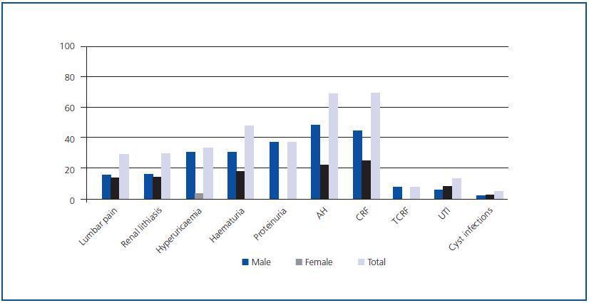
As an isolated manifestation, AH was the most frequent disorder and the initial sign of the disease in 68.42% of the patients, predominantly in men with respect to women (p < 0.05), followed by chronic renal failure (CRF), present in 68.29% of patients with ADPKD (Figure 1).

No statistically significant differences were found in the prevalence of the different initial clinical renal manifestations by gender, with the exception of AH, proteinuria, hyperuricaemia and renal lithiasis in which the prevalence was greater in men (p < 0.05) than in women. Quantification of proteinuria at the time of diagnosis was always less than 300mg/24 h.

Clinical renal manifestations during evolution

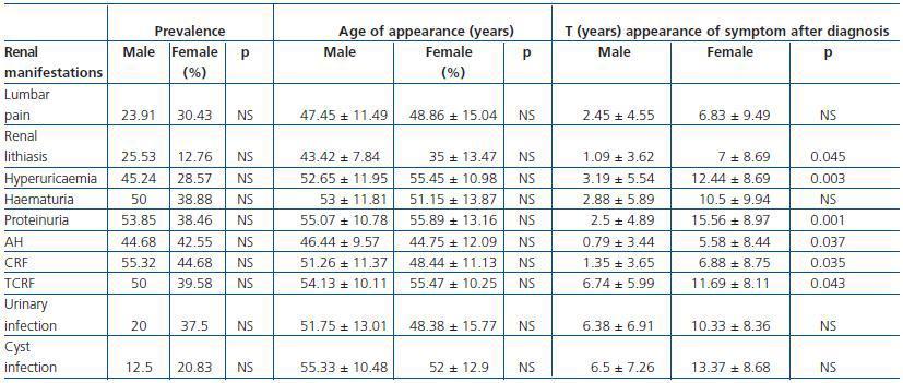
The most frequent renal manifestation during evolution was CRF, present in 100% of the studied patients, followed by proteinuria (92.31%) and terminal chronic renal failure (TCRF) (89.58 %) (Table 1). AH was present in 87.23% of the sick patients, statistically significant in the 30-60 age group (p < 0.01). The prevalence of renal manifestations during evolution showed no significant differences between men and women (Table 1).

Although the prevalence of urinary tract infections was higher in women, it had no statistical significance. Enterobacteria were responsible for 94.12% of the episodes and Escherichia coli was the most frequently isolated aetiological agent (70.59%).

The mean age at CRF onset was 50.02 ± 11.21 years, and that of TCRF 54.74 ± 10.07 years, independently of sex (p > 0.05) (Table 1). Patients diagnosed with CRF were found predominantly in the 30-60 age group (p < 0.05), as were patients with TCRF, in whom the difference was not statistically significant. When the disease was transmitted by the father, the mean age of onset of CRF and of TCRF was lower (p < 0.01).

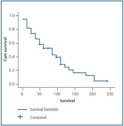
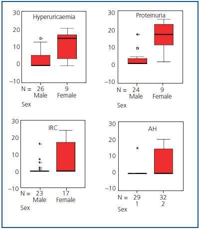
The evolution time to CRF and TCRF (Figure 2) from diagnosis of the disease was shorter in men than in women, as was the elapsed time from diagnosis of CRF to the start of renal replacement therapy (p < 0.05) (Table 1 and Figure 3). Patients with AH were more likely to exhibit CRF (p < 0.05).

Of all the patients who started renal replacement therapy, 83.72% were hypertensive and 13.95% normotensive. Patients with AH, proteinuria, macroscopic haematuria, lithiasis, abdominal pain, urinary tract infections, hepatic cysts and cyst infections were more likely to exhibit TCRF (p < 0.01). However, the probability of developing TCRF did not increase in patients with diverticula, macroscopic haematuria before 30 years of age or women with more than 3 children (p > 0.05).

No significant differences were found by sex in the age of onset of any of the renal manifestations (Table 1).

In addition to CRF and TCRF, the time of evolution from disease diagnosis to the appearance of hyperuricaemia, proteinuria, renal lithiasis and AH was also less in men than in women (p < 0.05) (Table 1 and Figure 3). No significant differences were found in the evolution period from diagnosis of the disease to the appearance of the other renal manifestations.

The recurrence of renal lithiasis was statistically significant in men.

Extrarenal manifestations

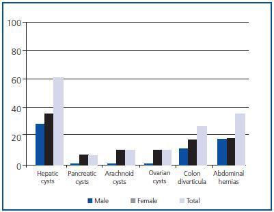
Hepatic cysts were the most frequent extrarenal manifestation, present at the time of diagnosis in 59.09% of patients, with greater prevalence in women (p < 0.05) (Figure 4). No statistically significant differences by sex were found in the prevalence of the other extrarenal manifestations diagnosed.

Survival and morbidity and mortality

Patients with ADPKD had a total of 6.17 ± 5.81 hospital admissions in our centre during the length of the study, 0.94 ± 0.63 admissions/year, with an average stay of 11.27 ± 4.91 days. No significant differences by sex were found in survival, or in the number of hospital admissions per year, nor in the average stay (Table 2).

Of the total number of patients with ADPKD, 39.58% died during the study, 29.17% men and 10.42% women (p < 0.05; RR = 2.3). Patient survival from the time of diagnosis was 13.4 ± 7.79 years, but it was not shown that AH, macroscopic haematuria, abdominal pain, cyst infections, renal lithiasis, urinary infections, the presence of hepatic cysts or a large number of childbirths were risk factors for mortality (p > 0.05).

The principal causes of death were of cardiovascular (42.1%) and infectious (42.1%) origin.

DISCUSSION

CLINICAL DIAGNOSIS

ADPKD is diagnosed in the majority of patients by characteristic clinical manifestations which usually appear in the third or fourth decade of life. In our study, the mean age of onset of the first clinical manifestations of ADPKD was earlier in women than in men. Studies published to date have not analysed the existence of differences by sex in age at disease diagnosis or in the prevalence of the majority of renal and extrarenal manifestations.1,4,7,9,12

In this series, the most frequent initial manifestation at diagnosis was AH, present in 68.42% of patients, with predominance in males and with a mean age of onset (45.77 ± 10.48 years) later than that described in the literature, with a mean age of 30 years;13,14 nevertheless, in our study it preceded measurable changes in renal function by several years.1,13,15 AH is, according to different publications, one of the guiding signs for ADPKD diagnosis, which is confirmed by this study. The fact that AH diagnosis in our population was later than that published1,14,16 could be explained by the current saturation of general medical consultations and by the lack of protocolised studies which clarify AH aetiology when it is light or easy to control; cases of ADPKD would thus be disguised among those in which early and vigorous treatment of complications would delay the onset of CRF and the progression of TCRF. The high prevalence in our study of CRF and TCRF secondary to ADPKD is in accordance with the ways the disease presents. During the evolution of ADPKD, AH was present in 87.23% of our patients, with no differences observed between the sexes. Evolution time from disease diagnosis to the appearance of AH was less in men. Its role in the progression of renal failure,13,17 already described, was demonstrated; we were unable to confirm this in patients under 35 given the low prevalence of young patients in our study. It has been proved, both in the general population and in sufferers of ADPKD that the prevalence of AH is significantly greater in men up to 45 years of age, at which point, although still prevalent in men, the differences are no longer significant. The shorter evolution time from disease diagnosis to AH onset in men may be due to the existence of cardiovascular risk factors (smoking habit, dyslipidaemia, central obesity) that favour the development of AH and that predominate in males between the ages of 20 and 55. From there on, hormonal influences, dyslipidaemia or central obesity cause these factors to increase significantly in women and, as a result, so does AH.14

In this series, proteinuria was one of the most frequent renal manifestations of ADPKD at the time of diagnosis, following AH, renal failure and haematuria. It was the presenting sign of the disease in 36% of the men, statistically significant with respect to the women, and the elapsed time from disease diagnosis to onset of proteinuria was shorter in men. AH and proteinuria present at the time of diagnosis has been related to worse renal prognosis and increased cardiovascular risk in ADPKD.17,18 Given that AH is correlated to the onset of proteinuria in various publications,19 the greater prevalence of AH in men at the time of diagnosis in this study would also explain the greater prevalence of proteinuria. Furthermore, AH, dyslipidaemia, central obesity and tobacco habit increase the risk of proteinuria and predominate in middle-aged men. During disease evolution, proteinuria becomes the second most frequent renal manifestation (92.31%) with no gender difference, which would explain the high percentage of patients with established kidney damage. The quantification of proteinuria was always lower than 300mg/dL, which agrees with that already described19 and there were no cases of nephrotic-range proteinuria. In the ADPKD patients with proteinuria studied, it was shown that they were more likely to present TCRF, in relation to secondary glomerular hyperfiltration.

Haematuria was a symptom presenting the disease in 47.06% of the cases, and was present in the evolution of the disease in 88.89% of our ADPKD patients. This prevalence was superior to that reported in literature, where the figure is 35-50%.1 Haematuria could be due to renal calculus, cyst infection or malignant tumours. The elevated prevalence of renal lithiasis and cyst infections in our series explains the higher prevalence of haematuria than that described. The underlying association of macroscopic haematuria with the progression of renal failure17,20 was evident, but that role was not demonstrated in patients under 30 probably due to the low prevalence of young patients with ADPKD. This deleterious effect of haematuria on kidney function can be explained both by the obstructive episodes secondary to clot formation and the hypovolaemic situations that it provokes.

The prevalence of urinary tract infections in our study was similar to that described by others. As in the general population, because the female urethra is shorter, prevalence was higher in women, but was not statistically significant.12,16E. coli was the most frequently isolated aetiological agent. The most frequent infections were cyst infections and acute pyelonephritis, with a determinant role in the progression of renal failure, probably in relation to the deterioration of kidney function secondary to episodes of haemodynamic instability derived from severe infection.

CRF was the second most frequent clinical manifestation at diagnosis and that of greatest prevalence in the evolution of the disease in our study, while in other series it is estimated that only around half of the patients with mutation will develop the disease before the sixth decade of life.15 Also, the need for renal replacement treatment in our work was higher than that described.18 The mean age of onset of CRF (50.02 ± 11.21 years) and TCRF (54.74 ± 10.07 years) was similar to that described in the literature for patients with PKD1 gene mutation and requiring renal replacement treatment (54.3 years),3,4 which leads us to assume a greater prevalence of patients in our population with PKD1 gene mutation if we limit ourselves to a clinical analysis. The age of onset of TCRF was earlier when transmitted by the father than by the mother and evolution time to CRF and TCRF from disease diagnosis was shorter in men than in women, a fact already reflected in different studies.3,21 In this study, men had more probability of rapid progression to renal failure, which appears to be related to a direct effect of the sexual hormones.21,22 Another sign of poor prognosis in the early evolution to both CRF and TCRF was the disease being inherited from the father.

As far as extrarenal manifestations,8 these predominated in women, although only hepatic cysts were statistically significant: the greater prevalence of these in women has been associated with the number of pregnancies, the taking of contraceptive pills and replacement hormonal therapy. Although there are contradictory results, we confirm that people with hepatic cysts have worse renal function than those who do not,3,16 probably due to the prejudicial effect that repeated infection in hepatic cysts would have on renal function.

In this study, a similar prevalence was observed in deaths of cardiovascular (42.1%) and infectious (42.1%) origin. Before 1975, the most frequent cause of death in ADPKD patients was infection (30%), uraemia (28%) and cardiovascular disease (21%). With the development of dialysis, the main cause of mortality has become cardiovascular (34%) followed by infections (20.4%).23 The principal cause of death of patients in renal replacement treatment and those subjected to renal transplant is cardiovascular; nevertheless, in our study there was a markedly high prevalence of deaths of infectious origin, which could be attributed to the immunodeficiency situation derived from a population with very high prevalence of TCRF secondary to ADPKD. The death rate was higher in men, but no significant differences were found in survival.

In spite of the mean age of ADPKD diagnosis in this study being lower in women, evolution up to the appearance of renal lithiasis, hyperuricaemia, proteinuria, AH and TCRF was shorter in men. This, together with the greater prevalence in men, at diagnosis, of risk factors involved in the progression towards TCRF, determined a more torpid evolution of the disease in men, an earlier start of renal replacement treatment and, consequently, greater mortality. In women, however, the evolution time of the disease was longer, which translated to a higher number of hospital admissions. We may think that in women morbidity predominates and in men, mortality, and it is probably not a reflection of the disease but an observation common to the entire population.

In short, in our study, male carriers of ADPKD were diagnosed later than women, which could explain their worse clinical evolution. In this population, AH was the most frequent clinical manifestation at diagnosis, and this classification should, therefore, be included in all cases with unknown AH. Also, infectious and cardiovascular complications were the principal cause of mortality, implying that infectious complications are a sign of alert in all ADPKD patients, including those not subjected to transplant.

Figure 1.

Table 1. Prevalence, age of appearance and elapsed time from disease diagnosis to the appearance of the different renal clinical manifestations by sex

Figure 2.

Figure 3.

Table 2. Prevalence and statistical significance by sex of deceased patients, mean age at time of death, survival since diagnosis, hospital admissions and mean stay

Figure 4.

Bibliography
[1]
1.  Bleyer AJ, Hart TC, Wilson PD. Polycistic kidney disease. N Engl J Med 2004;350:2622. [Pubmed]
[2]
2.  Daoust MC, Reynolds DM, Bichet DG, Somlo S. Evidence for a third genetic locus for autosomal dominant polycystic kidney disease. Genomics 1995;25:733-6. [Pubmed]
[3]
3.  Brown JA. End stage autosomal dominant polycystic kidney disease. N Engl J Med 2002;347:1504. [Pubmed]
[4]
4.  Igarashi P, Somlo S. Polycystic kidney disease. J Am Soc Nephrol 2007;18:1371-3. [Pubmed]
[5]
5.  Romão EA, Moysés Neto M, Teixeira SR, Muglia VF, Vieira-Neto OM, Dantas M. Renal and extrarenal manifestations of autosomal dominant polycystic kidney disease. Braz J Med Biol Res 2006;39:533-8. [Pubmed]
[6]
6.  Demetriou K, Tziakouri C, Anninou K, Eleftheriou A, Koptides M, Nicolaou A, et al. Autosomal dominant polycystic kidney disease-type
[7]
Ultrasound, genetic and clinical correlations. Nephrol Dial Transplant 2000;15:205-11. [Pubmed]
[8]
7.  Iglesias CG, Torres VE, Offord KP, Holley KE, Beard CM, Kurland LT. Epidemiology of adult polycystic kidney disease. Am J Kidney Dis 1983;2:630-9. [Pubmed]
[9]
8.  Peces R, Venegas JL. Quistes de las vesículas seminales e infertilidad en la poliquistosis renal autosómica dominante. Nefrologia 2005;25:78-80. [Pubmed]
[10]
9.  Nicolau C, Torra R, Badenas C, Vilana R, Bianchi L, Gilabert R, et al. Autosomal dominant polycystic kidney disease types 1 and 2: assessment of US sensitivity for diagnosis. Radiology 1999;213:273-6. [Pubmed]
[11]
Darnell A, Torra A, Badenas C, Pérez-Oller L, San Millán JL, Tellería D, et al. Estudio mutacional de los genes PKD1 y PKD2 (poliquistosis renal autosómica dominante). Nefrologia 2000;20:39-46. [Pubmed]
[12]
Torres MJ, Rodríguez JC, Hernández CR, Anabitarte A, Caballero A, Vázquez C, et al. Diagnóstico molecular de la poliquistosis renal autosómica dominante en la Comunidad Autónoma de Canarias. Nefrologia 2006;26:666-72. [Pubmed]
[13]
Torres VE. Extrarenal manifestations of autosomal dominant polycystic kidney disease. Am J Kidney 1999;34:45-8.
[14]
Ecder T, Schrier RW. Hypertension in autosomal-dominant polycystic kidney disease: early occurrence and unique aspects. J Am Soc Nephrol 2001;12:194-200. [Pubmed]
[15]
Kelleher CL, McFann KK, Johnson AM, Schrier RW. Characteristics of hypertension in young adults with autosomal dominant polycystic kidney disease compared with general US population.  Am J Hypertens 2004;17:1029-34. [Pubmed]
[16]
Chapman AB. Autosomal dominant polycystic kidney disease: time for a change. J Am Soc Nephrol 2007;18:1399-407. [Pubmed]
[17]
Gabow PA. Autosomal dominant polycystic kidney disease. More than a renal disease. Am J Kidney Disease 1990;16:403.
[18]
Choukroun G, Itakura Y, Albouza G, Christophe JL, Man NK, Grünfeld JP, et al. Factors influencing progression of renal failure in autosomal dominant polycystic kidney disease. J Am Soc Nephrol 1995;6:1634-42. [Pubmed]
[19]
Torres VE, Harris PC, Pirson Y. Autosomal dominant polycystic kidney disease. Lancet 2007;369:1287-301. [Pubmed]
[20]
Sharp CK, Gabow P. Factors relating to urinary protein excretion in children with autosomal dominant polycystic kidney disease. J Am Soc Nephrol 1998;9:1908-14. [Pubmed]
[21]
Johnson AM, Gabow PA. Identification of patients with autosomal dominant polycystic kidney disease at highest risk for end-stage renal disease. J Am Soc Nephrol 1997;8:1560-7. [Pubmed]
[22]
Torra R, Badenas C, Darnell A, Nicolau C, Volpini V, Revert L, et al. Linkage, clinical features, and prognosis of autosomal dominant polycystic kidney disease types 1 and 2. J Am Soc Nephrol 1996;7:2142-51.
[23]
Burtey S, Lossi AM, Bayle J, Berland Y, Fontés M. Mutation screening of the PKD1 transcript by RT-PCR. J Med Genet 2002;39:422-9. [Pubmed]
[24]
Fick GM, Johnson AM, Hammond WS, Gabow PA. Causes of death in autosomal dominant polycystic kidney disease. J Am Soc Nephrol 1995;5:2048-56. [Pubmed]
Download PDF
Idiomas
Nefrología (English Edition)
Article options
Tools
es en

¿Es usted profesional sanitario apto para prescribir o dispensar medicamentos?

Are you a health professional able to prescribe or dispense drugs?