Atherothrombotic disease (ATD) is a progressive disorder and its most common clinical manifestations, acute myocardial infarction and stroke, are responsible for the highest morbidity and mortality rates in the Western world. Sudden death due to infarction with no prior symptoms occurs in 50% of men and 64% of women.1
One of the main tools to control the incidence of vascular disease is prevention. Formulas are available to estimate cardiovascular risk (Framingham risk score, etc.)2 They are based on epidemiological studies that determine the risk of suffering a cardiovascular event (fatal or non-fatal) within 5 to 10 years.3 While the available system to calculate cardiovascular risk is easily generalisable, and universal, it does present some difficulties. The main drawback is its low sensitivity for detecting individuals at a high risk to suffer a cardiovascular event. Large epidemiological studies show that 62% of subjects with a prior history of myocardial infarction present none or only one of the cardiovascular risk factors,4 which, undoubtedly, prevents them from being properly identified at a time to take effective preventive actions.
In recent years, there have been significant technological advances in medical imaging techniques, particularly in the field of vascular disease. Below, we describe the advantages, disadvantages and uses of imaging techniques in the general population and in chronic kidney disease (CKD) patients.
GENERAL POPULATION
Various methods have been used for the diagnosis and prevention of cardiovascular disease (CVD). Studies on the calcification of coronary arteries using helicoidal or multidetector computed tomography (CT) accurately measure coronary calcium content (coronary calcium score). However, this method is not useful in patients at a low cardiovascular risk according to traditional estimation methods.5 Although coronary CT has a high specificity to diagnose coronary atherosclerosis, and some studies suggest that it directly correlates to mortality over time,6 this is a costly technique that requires advanced technology, and therefore, it cannot be used for the early detection of ATD in the general population. More significantly, coronary calcium scores measure the presence of calcium deposition in the coronary arteries, and therefore, it fails to diagnose early stages of vascular disease, as ATD is a progressive vascular disorder, and calcification only occurs in its advanced stages.
Coronary arteriography has recently been complemented with the use of intravascular ultrasound7 techniques that offer a more precise diagnosis of coronary atherosclerosis. However, this method is painful and expensive, and therefore cannot be considered a good option for the early detection of ATD in the general population.
Carotid ultrasound has several advantages compared to the previously described methods, but the use of this technique for early diagnosis of atherosclerosis is not generalised. Ultrasound of supra-aortic trunks is used in patients with symptomatic atherosclerosis, particularly those who have suffered a stroke. As these patients have already suffered an event due to arterial disease, we can only provide a diagnosis or confirmation of a suspected diagnosis and establish the surgical indication, rather than implementing preventive therapeutic interventions.
Applying this technique in early diagnosis of ATD requires rigorous training of the technician and the specialist reading the properly acquired images in order to obtain accurate parameters for the early diagnosis of the disease.
Carotid intima-media thickness (cIMT) measured by carotid ultrasound was described by Pignoli et al in 1984 and validated by the same authors in 1986. They conclusively established a strong correlation between the thickness of the arterial wall, measured by ultrasound, with the histological findings in these patients.8 Since then, carotid ultrasound has been known as a technique with potential applications in the field of atherosclerosis, but 20 years later, it is not part of the routine clinical practice to diagnose vascular disease, mainly due to the high variability in the resulting parameters, highly dependent on the acquisition technique employed. The recommendations of the XIII European Stroke Conference in 2004 (Manheim IMT Consensus)9 pconstitute a definite step forward in generalising the proper technique for clinical practice.
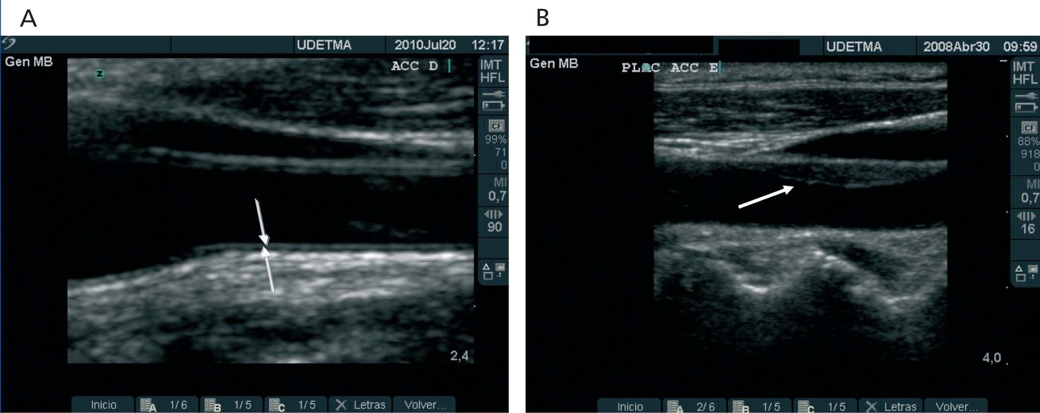
Carotid artery ultrasound, which measures cIMT and detects atheromatous plaques (Figure 1 A and Figure 1 B), is a strong indicator of overall vascular health. cIMT is a well recognized and accepted marker to predict cardiovascular disease.10
In clinical trials, the cIMT is used alongside traditional risk factors. These trials highlight the usefulness and importance of using non-invasive techniques to evaluate the effect of risk factors on the vascular wall. Through the years, clinical trials have provided evidence corroborating the importance of measuring cIMT to prevent cardiovascular events (the thicker the cIMT, the higher the risk of myocardial infarction and cerebral infarction).11
cIMT was accepted as a surrogate marker of atherosclerosis by the US Food and Drug Administration (FDA) and the European Agency for the Evaluation of Medicinal Products. At present, cIMT is the noninvasive technique most frequently used in clinical trials to determine atherosclerosis. Clinical guidelines and medical societies recommend it to obtain a more precise evaluation of cardiovascular health in selected populations,12 and in cardiovascular disease prevention programmes.13 Upon adjustment for age and sex, an increase of 0.1 mm in cIMT, indicates a 10%-15% increase in the risk of myocardial infarction, and a 13%-18% increase in the risk of stroke.11
Davidsson14 and coworkers have recently shown, in a multivariate analysis of 391 men in Sweden, odd ratios for cardiovascular events of 2.09 (95% confidence interval (CI) 1.05-4.16; p= 0.03) in subjects with atheromatous plaques in the carotid artery. Nambi et al15 published the latest results from the ARIC study, demonstrating a correlation between an increase in cIMT , or the presence of atheromatous plaques, and the incidence of cardiovascular events. They reported the predictive role of carotid ultrasound (cIMT and plaques) in a large epidemiological study of 13,145 healthy individuals, who presented 1,812 adverse cardiovascular events over a 15-year follow up. The area under the curve estimating the sensitivity and accuracy of their logistic regression analysis increased significantly when cIMT and the presence of atheromatous plaque were added to the list of conventional cardiovascular risk factors.
CAROTID ULTRASOUND IN CHRONIC KIDNEY DISEASE
Current tables to estimate cardiovascular risk have not been designed for CKD patients, whose main cardiovascular risk factors are different from those in the general population. CKD patients experience the so-called “reverse epidemiology”. In studies in haemodialysis patients, no association was found between serum cholesterol levels or systolic blood pressure with coronary artery disease, cerebrovascular disease or peripheral vascular disease.16,17 Furthermore, there are other “non-traditional” and “emerging” factors that play a role in the pathogenesis of atherosclerosis in CKD, such as anaemia, chronic inflammation, oxidative stress, and abnormalities in mineral metabolism that are not taken into account when using traditional predictors cardiovascular risk.
This evidence, along with the high incidence rate of CVD among CKD patients,18 supports the use of new, more sensitive tools for the early detection and prevention of cardiovascular disease.19 Guidelines from the European Society of Cardiology recommend a close monitoring of traditional cardiovascular risk factors in patients with chronic kidney disease,20 , Furthermore, the National Kidney Foundation (NKF) recommends the use of carotid ultrasound for a more precise assessment of cardiovascular health in kidney disease.21
Because cardiovascular disease is the main cause of morbidity and mortality in CKD,22 early diagnosis of vascular disease using ultrasound techniques is becoming increasingly widespread in this patient population.
We lack conclusive evidence on how changes in cIMT relate to cardiovascular risk factors in CKD, but it has been reported that cIMT increases as glomerular filtration rate decreases.23
Our group has confirmed the high prevalence and severity of atherosclerosis in patients with different stages of kidney disease (n=409).24 The diagnostic methods used were carotid ultrasound and the ankle-brachial index (ABI). Both tests have been validated as predictors of adverse cardiovascular events.25 We have developed a classification of vascular disease based on the results of ABI and carotid ultrasound, using for the cIMT reference intervals adjusted for age and sex . We found that a high percentage of kidney patients had atheromatous vascular disease, and this percentage was much higher than in age- and sex-matched control subjects with a normal kidney function (n=851). Relevant findings from this study were the higher prevalence of ATD in patients on dialysis compared to those at earlier CKD stages, and the lack of correlation between vascular disease and LDL cholesterol levels. The presence of atheromatous plaque was higher in dialysis patients (78.3%)26 compared to pre-dialysis patients (55.6%)27 or the normal control group (43.2%). Variables with a positive association with atherosclerosis included age, dialysis treatment, and serum levels of plasma C-reactive protein. Being a female was a protective factor also in CKD.
Carotid ultrasound also offers a critical additional information, the assessment of vascular calcification. Vascular calcification is a strong predictor of morbidity and mortality in the general population and in CKD In CKD patients, vascular calcification is more prevalent, appears at an earlier age, and is more severe.28 The degree of arterial calcification can be measured using CT to obtain the Agatston score, which is the only quantitative method currently available, and therefore it is the gold standard, mostly used in clinical research. Semi-quantitative methods have been proven useful, as those described by Adragao and Kauppila. They employ simple radiology, are more adapted to clinical practice, and have proved to have a good predictive power of cardiovascular events in CKD.
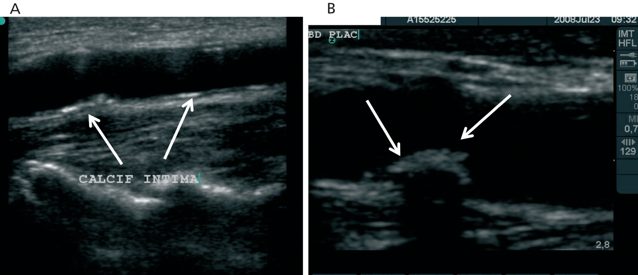
Carotid ultrasound is also the only available technique to localise the site of calcium deposition within the arterial wall. Our group has completed an ultrasound study examining the location of calcium in carotid and femoral artery wall..The brachial artery served as a negative control since this area is free of atherosclerosis.29 We observed that calcification was localized exclusively in the intimal layer, following two distinct patterns: 1) Calcification of the atheromatous plaque (Figure 2 A) and 2) Linear calcification of the intimal layer, anatomically unrelated to the atheromatous plaque (Figure 2 B). Multivariate analysis associated this previously unrecognized intimal calcium location with age, serum levels of phosphate and C-reactive protein, and with the presence of atheromatosis. These results underscore the contribution of inflammation and atheromatosis to the calcification of elastic arteries. Although serum phosphate levels remain a not fully resolved issue, we must start focussing on preventing atheromatosis and decreasing the pro-inflammatory status of CKD patients as much as possible.
CONCLUSIONS
Carotid ultrasound is a tool with a number of advantages compared to other methods. First, it is easy to use and does not require complex equipment. It can be performed in outpatient clinics or at the bedside; and is inexpensive and non invasive. Secondly, it provides doctors with valuable information for treatment, including the atheromatous burden, the severity of the calcification, and the location of calcium deposition within the vascular wall. Thirdly, it detects ATD at early stages, prior to the appearance of atheromatous plaque. Therefore, it constitutes an ideal technique to obtain evidence-based recommendations for ATD prevention.
At present, we lack studies addressing the prognostic value of either the calcification of atheromatous plaques (type V plaque according to the American Heart Association) or the new linear profile of intima calcification in kidney disease patients.
The Spanish study NEFRONA (www. nefrona.es) has been designed to answer important questions on the pathogenesis and progression of ATD at all CKD stages compared to age- and sex- matched controls with normal renal function through measurements of the distinct atherosclerosis and calcification patterns between groups using arterial ultrasound.
Conflicts of Interest
Authors declare potential Conflicts of Interest
Honoraria as speaker: Shire, Abbott, Genzyme and Amgen
Acknowledgements
Grants: FIS (Carlos III Health Institute).
Figure 1. Ultrasound image of carotid arteries
Figure 2. Calcification patterns depicted by ultrasound


