
Testosterone deficiency is a prevalent condition in male patients with chronic kidney disease. However, it is not known whether the type of renal replacement therapy has an impact on testosterone deficiency that accompanies loss of renal function.
MethodsThe cross-sectional study enrolled 79 prevalent male patients on dialysis; 43 on haemodialysis (HD) and 36 on peritoneal dialysis (PD). The median age was 69 years and 31.6% were diabetics. Endogenous testosterone levels were measured by immunoluminescence assay (normal range 3–10.5ng/ml), while nutritional/inflammatory markers, bone and mineral metabolism markers, anemia, type of dialysis technique and time on dialysis were also assessed. Body composition was evaluated by bioimpedance vector analysis and bioimpedance spectroscopy. Testosterone deficiency was defined as levels below 3ng/ml.
ResultsMean testosterone levels were 8.81±6.61ng/ml. Testosterone deficiency affected 39.5% of HD patients and only 5.6% of PD patients. In the univariate analysis, testosterone levels were directly correlated with type of dialysis technique (HD) (Rho Spearman 0.366; p<0.001) and time on dialysis (Rho −0.412; p=0.036) and only with the HD technique in the multivariate analysis. No other significant correlations were found.
ConclusionsCirculating testosterone levels in men on dialysis were independently associated with HD technique. It can be concluded that a new factor—namely the dialysis technique—may be associated with falling testosterone levels and the associated loss of muscle mass and inflammation. Further studies are needed to establish whether the dialysis technique itself triggers testosterone elimination.
Los varones con enfermedad renal crónica cursan a menudo con deficiencia en testosterona. Se desconoce si el déficit de testosterona que acompaña a la pérdida de función renal se asocia con el tipo de tratamiento sustitutivo de la función renal.
MétodosEl estudio de corte transversal incluyó 79 varones prevalentes en diálisis, 43 en hemodiálisis (HD) y 36 en diálisis peritoneal (DP). Con una edad media de 69 años, el 31,6% eran diabéticos. Se evaluaron los niveles de testosterona endógena (inmunoluminiscencia: N 3–10.5ng/ml), marcadores nutricionales/inflamatorios, marcadores de metabolismo óseo mineral, anemia, tipo de técnica y permanencia. La composición corporal fue estimada mediante bioimpedancia vectorial y espectroscópica. Se considera déficit de testosterona cuando los niveles son inferiores a 3ng/ml.
ResultadosLos niveles de testosterona medios fueron 8,81±6,61ng/ml. El 39,5% de los pacientes en HD y el 5,6% de los de DP presentaban déficit de testosterona. Los niveles de testosterona se correlacionaron directamente con el tipo de técnica, HD (rho Spearman 0,366; p < 0,001) y el tiempo de permanencia (Rho −0,412; p=0,036) en el análisis univariante y solo con la técnica de HD en el multivariante. No se encontraron otras correlaciones significativas.
ConclusionesLos niveles circulantes de testosterona en hombres en diálisis se asocian de manera independiente con la técnica de HD. Se puede concluir que, en la reducción de testosterona que acompaña de manera natural a la pérdida de masa muscular e inflamación, se asocia un nuevo factor que es la técnica dialítica. Se necesitan estudios para elucidar si la técnica per se favorece la eliminación de testosterona.
There are several mechanisms involved in the generation of testosterone deficit in males. In addition to a decrease in hormone production, there are other mechanisms that have to do with abnormalities of the hypothalamic-pituitary axis and also in relation with the storage and transport of the hormone. In the case of older patients with some type of hypogonadism, it is necessary to consider situations such as malnutrition and low serum protein concentration, since most of the circulating testosterone is bound to a protein and only a small proportion is circulating as a free form. Likewise, uremic toxicity could contribute to testosterone deficiency.
In chronic kidney disease (CKD) there is some degree of hypogonadism and androgen deficiency that is considered one common feature among renal patients. Progressive deterioration of renal function leads to gradual sexual dysfunction which does not appear to be corrected by dialysis. The deficit of testosterone correlates with the reduction of renal function. Testosterone is an anabolic hormone that plays an important role in muscle anabolism. Low testosterone level is associated with endothelial dysfunction and the possibility of cardiovascular events.1 Low testosterone in chronic renal male patients may be involved not only in reduction of libido and erectile dysfunction2 but also in cognitive impairment,3 anemia,4 cardiovascular disease5 and increased mortality.6
Aging aggravates testosterone deficiency that contributes to the development of atherosclerosis which is accentuated by renal failure. Low testosterone levels is thought to be present in 26–66% of patients with different stages of CKD.2 It is not known whether testosterone deficiency associated with renal failure correlates with muscle loss in men with advanced CKD. Previous studies have evaluated the testosterone deficiency in chronic hemodialysis (HD) patients and its relation with age, anemia, frequency of cardiovascular events and mortality; however in peritoneal dialysis (PD) patients there are not many studies evaluating serum testosterone levels and the potential impact on body composition, muscle strength and nutrition in general.
The aim of the present study is to evaluate testosterone levels in patients on PD and HD to assess the effect of the dialysis modality.
Patients and methodsThis is an observational study in a cohort of 79 prevalent male dialysis patients, stable for more than 6 months. A total of 43 patients were on HD for a period of 80.4±15 months and 36 patients were on PD for 32.6±27.7 months (p<0.001). Patients on HD were 68.84±13.22 years old and received HD for 12hours per week, none was on an on-line technique. Patients on PD were 60.5±15.6 years old and there were receiving with continuous ambulatory PD (n=13 (36.1%)) or with an automated technique (n=23 (63.9%)). Diabetic patients were 41.9% and 19.0% in HD and PD respectively (p=0.03). The adequacy of dialysis in HD patients was assessed monthly in a midweek session. In PD patients adequacy of dialysis was measured by weekly Kt/v and the residual renal function was assessed by urea and creatinine clearance. Four patients were excluded because they were on hormonal therapy for due to prostate neoplasia.
Endogenous testosterone level was measured by immunoluminescence (normal values were 3–10.5μg/ml). Testosterone deficiency was considered if serum level was <3ng/ml. Nutritional and inflammatory markers including serum albumin, prealbumin, protein catabolic rate (nPCR), and C-reactive protein (CRP) were also measured. Other parameters evaluated were serum concentration phosphorus, 25OH Vit D, intact parathyroid hormone (iPTH) and the hemoglobin level.
Body composition was assessed by vector bioimpedance analysis (BIVA) and spectroscopic bioimpedance (BIS). BIVA measurements were performed at the end of the mid-week session in HD patients, and in the early-morning in PD patients, with the abdomen filled with dialysis fluid. The BIVA was performed using an alternating sinusoidal current of 800A at 50kHz, with a standard total tetrapolar technique (EFG Impedance Analyzer; Akern, Florence, Italy). The test was performed under standardized conditions: a calm environment, with a temperature of 22–24°C, in supine position at the end of the HD session on the opposed side of the vascular access in the case of AVF, and in either side in the case of catheters.
The BIS was performed with a portable monitor for total body bioimpedance BCM (Fresenius Medical Care D, GmbH). Resistance and reactance were measured with the BCM at 50 different frequencies, covering a frequency spectrum from 5 to 1000kHz. Intracellular and extracellular resistances were determined.
The parameters studied by bioimpedance included the nutritional status, fat-free mass, lean body mass and muscle mass.
Statistical analysis was performed using the statistical software SPSS 21 (SPSS Inc, Chicago, IL, USA) for Windows. Age is expressed as median. Epidemiological data are expressed as mean±standard deviation. Statistical significance was considered with a p<0.05. Normality of the sample was assessed by the Kolmorogov–Smirnov test. Non-parametric categorical variables were assessed using the Chi-square test. Comparisons between groups with different testosterone levels were established by Student's t test for continuous variables with a normal distribution and by Mann–Whitney for continuous variables without normal distribution. Given the small sample size, Spearman's correlation was used in HD and DP to establish the relationship between testosterone levels and anthropometric parameters, nutritional markers, body composition parameters, markers of mineral and bone metabolism, inflammatory markers and the period of time on each technique. Univariate and multivariate analysis was used to establish the independent association between testosterone and two dependent variables: time on dialysis and dialysis technique.
Since the values of CRP are not normally distributed, the Ln was used in the multivariate analysis. Binary logistic regression analysis was performed using testosterone deficiency as dependent variable and the variables that were significant in the univariate analysis.
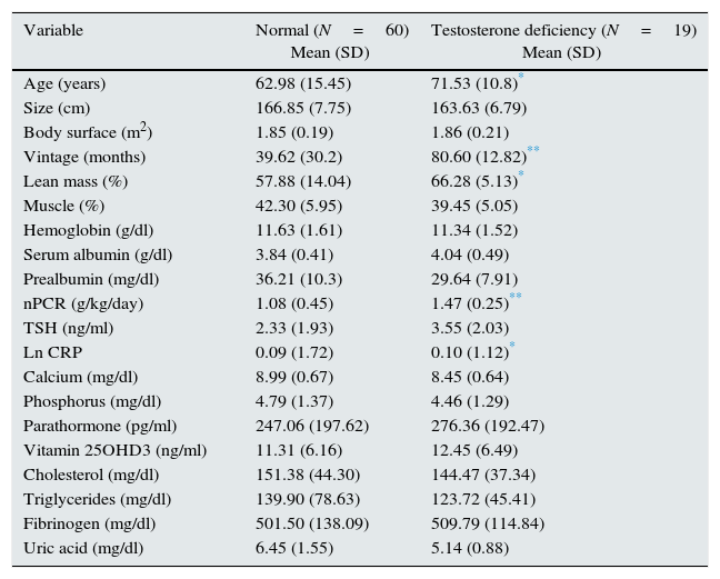
ResultsTestosterone deficiency was observed in 19 out of the 79 patients evaluated (21.4%); 17 patients were on HD and 2 on PD. The patients in HD were older than PD but the difference was not significant. The description of the study population is shown in Table 1. The mean level of testosterone in PD and HD patients were 10.84±4.67 versus 7.18±6.7ng/ml respectively (p<0.001). In general as the tertiles of testosterone decreased, the patients were older. Normal testosterone levels were observed in the younger patients and the lowest testosterone levels were observed among the oldest patients (data not shown). Testosterone values were lower in diabetics than in nondiabetics (6.58±0.55 vs. 9.9±6.6ng/ml, p=0.024). Table 1 shows the values of the parameters studied in all patients (HD+PD) with testosterone levels above and below 3ng/ml. As expected, significant differences were observed in age and dialysis vintage, although without statistical differences if patients were separated according to the dialysis technique. Residual renal function did not show an effect on testosterone levels.
Descriptive statistics of 79 patients (HD, n=43 and DP, n=36) separated according to the presence of testosterone deficiency.
| Variable | Normal (N=60) Mean (SD) | Testosterone deficiency (N=19) Mean (SD) |
|---|---|---|
| Age (years) | 62.98 (15.45) | 71.53 (10.8)* |
| Size (cm) | 166.85 (7.75) | 163.63 (6.79) |
| Body surface (m2) | 1.85 (0.19) | 1.86 (0.21) |
| Vintage (months) | 39.62 (30.2) | 80.60 (12.82)** |
| Lean mass (%) | 57.88 (14.04) | 66.28 (5.13)* |
| Muscle (%) | 42.30 (5.95) | 39.45 (5.05) |
| Hemoglobin (g/dl) | 11.63 (1.61) | 11.34 (1.52) |
| Serum albumin (g/dl) | 3.84 (0.41) | 4.04 (0.49) |
| Prealbumin (mg/dl) | 36.21 (10.3) | 29.64 (7.91) |
| nPCR (g/kg/day) | 1.08 (0.45) | 1.47 (0.25)** |
| TSH (ng/ml) | 2.33 (1.93) | 3.55 (2.03) |
| Ln CRP | 0.09 (1.72) | 0.10 (1.12)* |
| Calcium (mg/dl) | 8.99 (0.67) | 8.45 (0.64) |
| Phosphorus (mg/dl) | 4.79 (1.37) | 4.46 (1.29) |
| Parathormone (pg/ml) | 247.06 (197.62) | 276.36 (192.47) |
| Vitamin 25OHD3 (ng/ml) | 11.31 (6.16) | 12.45 (6.49) |
| Cholesterol (mg/dl) | 151.38 (44.30) | 144.47 (37.34) |
| Triglycerides (mg/dl) | 139.90 (78.63) | 123.72 (45.41) |
| Fibrinogen (mg/dl) | 501.50 (138.09) | 509.79 (114.84) |
| Uric acid (mg/dl) | 6.45 (1.55) | 5.14 (0.88) |
nPCR: normalized protein catabolic rate; CRP: C reactive protein.
Hemoglobin values were similar in patients with low and normal testosterone level. Among the nutritional parameters, only prealbumin was lower in patients with testosterone deficiency, whereas values of serum albumin and nPCR were higher in patients with hormone deficiency. C-reactive protein was higher in patients with normal testosterone values; if HD and PD patients are analyzed separately this difference is maintained only in HD patients. Phosphorus and iPTH do not appear to be different in patients with testosterone deficiency, and vitamin D is low in all patients, with or without testosterone deficiency. Data obtained by bioimpedance showed differences according to the testosterone levels. With a similar body surface, patients with testosterone deficiency had significantly higher values of lean mass (Table 1), while there were no differences in percentages of fat and muscle mass. In a separate analysis of patients on HD and PD no differences were observed in any of the parameters measured by bioimpedance. There was no significant difference in testosterone levels between the 2 techniques of DP (DPCA and DPA) (12.29±4.9 vs. 10.07±4.43ng/ml). The chi-square test and Fisher's test compared testosterone levels (0 normal/1 deficit) with diabetic status and dialysis techniques. Only the HD technique was significantly associated to testosterone deficiency (p<0.001). In the binary logistic regression testosterone deficiency was significantly associated with dialysis vintage (OR: 1.057; 95% CI: 1.004–1.13); and the HD technique (OR: 11.15; CI: 2355–52,453).
DiscussionTestosterone deficiency has negative effect on the health of dialysis patients, and the associated sexual dysfunction has negative impact on their quality of life.7 Our study in male patients in HD and PD reveals that a significant percentage of patients present this hormonal deficit. This was previously observed in HD patients8 and in another work that included patients in the both HD and PD, but levels of testosterone were not compared between these two techniques.9 An important finding of our study is that patients with PD have higher testosterone levels than HD, with a very small percentage of PD patients showing values below normal. Therefore, to date, the present study is the only one that shows the influence of a renal replacement technique in the development of hypogonadism. We observed that classic factors associated to testosterone deficit in the general population, such as age3 and diabetes mellitus,5 have the same significance in dialysis patients. However by multivariate analysis differences in testosterone levels between the two techniques are not justified by differences in age or percent of diabetic patients. Our data indicate that patients with a longer period of time on dialysis have lower testosterone levels but, again, the differences in testosterone between the two dialysis techniques cannot be justified by the time on renal replacement therapy. A question to be addressed is whether the dialysis technique may condition testosterone removal through the dialysate or the effluent as an important reason for the observed differences between HD and DP; however, there is not research done evaluating the loss of testosterone by the two techniques. One study performed in only HD patients10 evaluates the amount of the hormone removed in a 4-h HD session, but it is unknown how it compares with testosterone clearance by PD. Our results may be justified if the removal of testosterone is less in PD than HD. Another possible explanation may be that more peritoneal protein losses during PD could produce a greater elimination of the protein-bound testosterone, which would increase the ratio free testosterone to protein-bound of testosterone resulting in more free testosterone.
The reported relationship of testosterone deficiency and the development of anemia and the lower response to erythropoietic factors in patients on HD2,4,6 is not confirmed in our patients since hemoglobin levels are similar in patients with and without testosterone deficiency.
The results obtained on the body composition with the use of bioimpedance show a relationship between testosterone deficiency and a higher percentage of lean body mass; interestingly, this is accompanied by greater protein intake and a tendency to have higher levels of albumin. Certainly, it would not be appropriate to say that these finding are related with an effect of low testosterone on appetite. In this regard, a recent study11 shows that androgen deficiency decreases of lean body mass, muscle size and strength and increases in fat mass. Patients receiving daily supplement of testosterone there was a reduction in body fact. In our study, with only 2 patients with testosterone deficiency in PD, it is very difficult to assess statistical differences between HD and DP. According to our results, neither vitamin D nor PTHi appear to be associated to testosterone levels. Significantly higher C-reactive protein level in patients with normal testosterone levels seem to correlate with a higher degree of inflammation in HD patients. Inflammation is common in patients with CKD and, especially in those undergoing replacement therapy; the uremic phenotype is associated with a state of immunosuppression that affects both innate and adaptive immunity. The end result is that patients on dialysis have inflammation with increased susceptibility to infections. In our series we have not been able to rule out the causes of inflammation and it does not appear that testosterone deficiency per se is a cause or a consequence of inflammation.12
Given the negative effect of testosterone deficiency in CKD patients which may even influence mortality13 treatment with hormonal supplements may be considered.3 There is clinical experience with testosterone supplements in patients with heart failure14 and in HD patients.10 Treatment with testosterone is not without risk, especially in the elderly, which is the case in among dialysis patients. Liver toxicity, risk of prostate cancer, and volume retention are some of the side effects of testosterone administration.15 The use of testosterone supplementation is considered safe in general, but in CKD patients it needs further evidence.
The measurement of the amount of hormone eliminated with PD, not performed in our study, may have been useful to determine whether PD helps to maintain higher testosterone levels, that has important clinical implications.
Our study has two important limitations; therefore the results must be interpreted with caution. One limitation is to the size of the sample, which is insufficient and decisively claim that the HD technique and the period of time on dialysis are responsible for the testosterone deficiency. Another limitation is the technical impossibility to determine testosterone levels in both the dialysate fluid and in peritoneal effluent.
In conclusion, dialysis patients have a testosterone deficit that could have adverse effects. Most PD patients present normal levels of the hormone as compared to the lower values in patients on HD. Patients on HD and those with longer time on replacement treatment have a higher risk of testosterone deficiency. Differences between PD and HD testosterone losses deserve further investigation.
Conflicts of interestThe authors have no conflicts of interest to declare.
Please cite this article as: Cigarrán S, Coronel F, Florit E, Calviño J, Villa J, Gonzalez Tabares L, Herrero JA, Carrero JJ. Déficit de testosterona en los pacientes en diálisis: diferencias según la técnica de diálisis. Nefrologia. 2017;37:526–530.


