Journal Information
Vol. 36. Issue. 4.July - August 2016
Pages 333-464
Visits
14744
Vol. 36. Issue. 4.July - August 2016
Pages 333-464
Review
Open Access
Bone mineral disorder in chronic kidney disease: Klotho and FGF23; cardiovascular implications
Enfermedad óseo mineral relacionada con la enfermedad renal crónica: Klotho y FGF23; implicaciones cardiovasculares
Visits
14744
Laura Salanova Villanueva
Corresponding author
aelita.sv@gmail.com

Corresponding author.
, Carmen Sánchez González, José Antonio Sánchez Tomero, Abelardo Aguilera, Esther Ortega Junco
Servicio de Nefrología, Hospital de La Princesa, Madrid, Spain
This item has received

Under a Creative Commons license
Article information
Abstract
Full Text
Bibliography
Download PDF
Statistics
Figures (2)
Tables (1)
Table 1. Regulation of bone mineral disorder related to chronic kidney disease.
Abstract

Cardiovascular factors are one of the main causes of morbidity and mortality in patients with chronic kidney disease. Bone mineral metabolism disorders and inflammation are pathological conditions that involve increased cardiovascular risk in chronic kidney disease. The cardiovascular risk involvement of bone mineral metabolism classical biochemical parameters such as phosphorus, calcium, vitamin D and PTH is well known. The newest markers, FGF23 and klotho, could also be implicated in cardiovascular disease.

Keywords:
Cardiovascular risk
Chronic kidney disease
Klotho
FGF23
Resumen

Una de las principales causas de morbimortalidad en el paciente con enfermedad renal crónica es la cardiovascular. La inflamación y las alteraciones en el metabolismo óseo mineral son una condición patológica que conlleva aumento del riesgo cardiovascular en la enfermedad renal crónica. Los parámetros bioquímicos clásicos del metabolismo óseo mineral como fósforo, calcio, vitamina D y PTH tienen una implicación muy conocida en el riesgo cardiovascular. Los nuevos marcadores, FGF23 y klotho, también podrían estar implicados en la enfermedad cardiovascular.

Palabras clave:
Riesgo cardiovascular
Enfermedad renal crónica
Klotho
FGF23
Full Text
Introduction

Chronic kidney disease (CKD) is a public health problem that is on the rise, with an ever-increasing incidence and prevalence (10% of the general population).1,2 The main cause of mortality in the CKD patient is cardiovascular (CV),3 with an increase in risk of up to 20 times that of the general population, even in initial stages.4–6 Up to 80% of patients with CKD have associated CV disease: hypertension (36%), ischaemic cardiomyopathy (22–39%), atrial fibrillation (30%), valvular heart disease (24%) and left ventricular hypertrophy (LVH) (50–75% in stage 3–4 CKD).7,8

Cardiovascular risk factors in the chronic kidney disease patient

The greater cardiovascular risk (CVR) of patients with CKD is explained by the high presence of classic risk factors and the overlapping of specific factors of a uraemic state as well as by the inflammatory state of CKD; stage 5 adds other factors related to dialysis or transplant that causes an excess of vascular calcification9,10 (Fig. 1). Bone mineral disorder related to CKD (BMD-CKD) plays a crucial role in CKD morbidity and mortality. BMD-CKD includes clinical-chemistry and skeletal abnormalities and extraskeletal calcifications caused by abnormalities in mineral metabolism in CKD. CKD-MBD I presents as one or a combination of the following manifestations11:

  • Abnormalities in calcium (Ca), phosphate (P), parathyroid hormone (PTH) and vitamin D, klotho, or fibroblast growth factor 23 (FGF23).

  • Abnormalities in skeletal remodelling, mineralisation, volume, growth or fragility.

  • Calcifications of cardiovascular or other soft tissues.

Fig. 1.

CVR factors involved in renal and cardiac impairment.

(0.24MB).

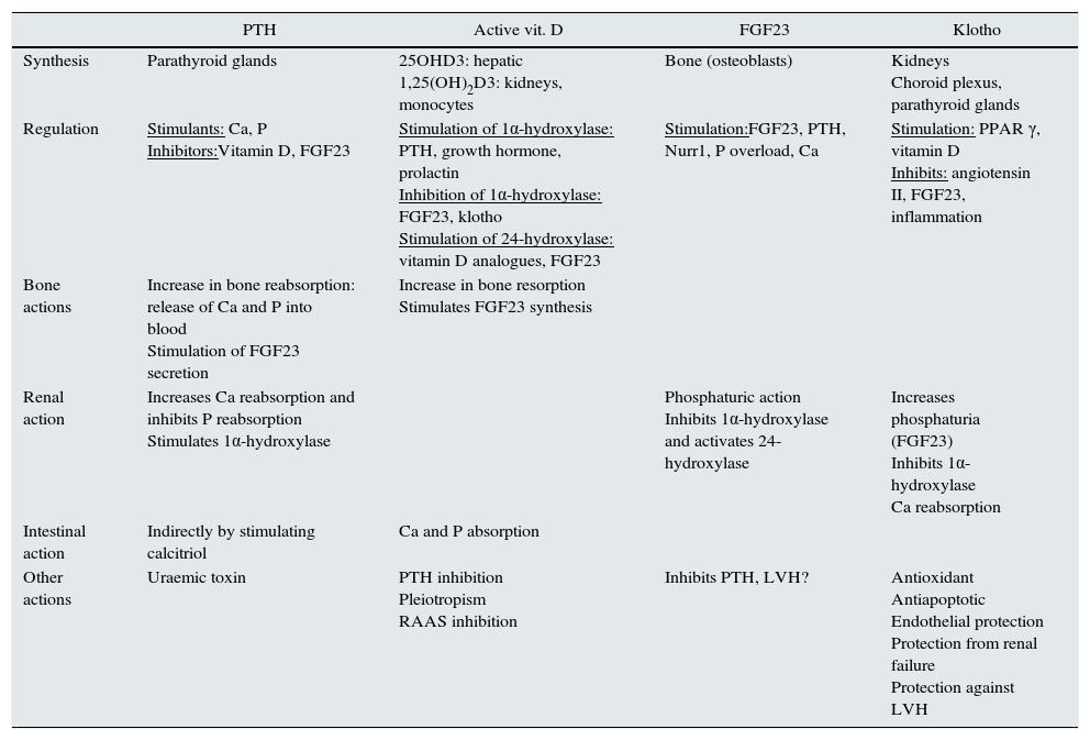
Studying markers of BMD-CKD sometimes allows CVR to be predicted. Table 1 summarises the role of the factors involved in BMD-CKD. The role of klotho and FGF23 in CVD is reviewed below.

Table 1.

Regulation of bone mineral disorder related to chronic kidney disease.

  PTH  Active vit. D  FGF23  Klotho 
Synthesis  Parathyroid glands  25OHD3: hepatic
1,25(OH)2D3: kidneys, monocytes 
Bone (osteoblasts)  Kidneys
Choroid plexus, parathyroid glands 
Regulation  Stimulants: Ca, P
Inhibitors:Vitamin D, FGF23 
Stimulation of 1α-hydroxylase: PTH, growth hormone, prolactin
Inhibition of 1α-hydroxylase: FGF23, klotho
Stimulation of 24-hydroxylase: vitamin D analogues, FGF23 
Stimulation:FGF23, PTH, Nurr1, P overload, Ca  Stimulation: PPAR γ, vitamin D
Inhibits: angiotensin II, FGF23, inflammation 
Bone actions  Increase in bone reabsorption: release of Ca and P into blood
Stimulation of FGF23 secretion 
Increase in bone resorption
Stimulates FGF23 synthesis 
   
Renal action  Increases Ca reabsorption and inhibits P reabsorption
Stimulates 1α-hydroxylase 
  Phosphaturic action
Inhibits 1α-hydroxylase and activates 24-hydroxylase 
Increases phosphaturia (FGF23)
Inhibits 1α-hydroxylase
Ca reabsorption 
Intestinal action  Indirectly by stimulating calcitriol  Ca and P absorption     
Other actions  Uraemic toxin  PTH inhibition
Pleiotropism
RAAS inhibition 
Inhibits PTH, LVH?  Antioxidant
Antiapoptotic
Endothelial protection
Protection from renal failure
Protection against LVH 
FGF23

This is a 32-kDa, 251-amino acid protein synthesised and secreted by bone cells, mainly osteocytes. It belongs to the “phosphatonin” hormone group12 owing to its involvement in renal phosphorus elimination. It has been pathophysiologically linked to the so-called “rare hypophosphataemic syndromes”,13 characterised by mineralisation defects and bone deformities, hypophosphataemia, renal P loss and inappropriately low calcitriol levels. It is considered to be one of the main factors in P metabolism regulation.14–16 The biological action of FGF23 depends on the klotho gene14,17–19 which acts as its co-receptor. FGF23 is also expressed in the heart, liver, thyroid gland, parathyroid glands, intestine and skeletal muscle.19

FGF23 regulation

FGF23 regulation is determined by:

  • 1.

    Active vitamin D: calcitriol directly and indirectly increases FGF23 transcription through extracellular signalling pathways mediated by leptin and interleukin 6.20 Calcitriol also increases expression of the nuclear receptor associated with protein 1 (Nurr1) in bone cells and of PTH, which leads to an increase in FGF23.21

  • 2.

    Ca levels: high calcium stimulate FGF23 secretion.22,23

  • 3.

    Hyperparathyroidism: the increase in PTH linked to CKD may stimulate FGF23 secretion through Nurr1.19,21 In primary hyperparathyroidism (PHPTH), FGF23 would hypothetically be suppressed since PTH hypersecretion would cause hypophosphataemia so one important stimuli of FGF23 secretion would be reduced.24 However, experimental studies have demonstrated that mice with PHPTH have higher FGF23 levels than controls and that PTH directly stimulate FGF23,25 since FGF23 decreases following parathyroidectomy.25 Yamashita et al.24 demonstrated in patients with PHPTH that FGF23 levels were high compared to healthy controls. However, in this same study, patients without CKD or PHPTH had no differences in FGF23 levels compared to controls,24 and the authors concluded that in PHPTH renal function is a determining factor in FGF23 levels.24

  • 4.

    P levels: serum P levels have been positively correlated with FGF23 concentration in patients with CKD.26 However, restricting dietary P in patients with CKD has shown contradictory results in FGF23 levels. Some studies have demonstrated that restricting dietary P fails to cause a drop in FGF23 levels in patients with stage 3–4 CKD27 and does not change them in healthy volunteers.28 Despite this, a drop in P absorption with phosphate binders such as sevelamer causes a drop in FGF23 levels.29,30

  • 5.

    Reduced renal synthesis of klotho: FGF23's affinity for its receptor (especially FGFR1 in the kidneys) is very low.31 Under physiological conditions, when bound to FGFR1, FGF23 would be unable to generate signal transduction.31 In the presence of klotho, FGF23 affinity for its receptor FGFR1 increases significantly and allows its activation with physiological concentration of FGF23.31 A reduction of klotho expression could cause resistance to the action of FGF23; in CKD this resistance would lead to a reduction in the fractional excretion of P and an increase in serum P concentration which in turn stimulate FGF23 secretion.32

Interestingly, iron infusion and low iron levels may induce synthesis of FGF23, although not in its active form.22 Metabolic acidosis, oestrogens and leptin also cause increases in FGF23.33,34

Biological actions of FGF23

FGF23 has target receptors called FGFR1, FGFR3 and FGFR4 and the transmembrane receptor β-glucuronidase. To exert its action on FGFR1 in the kidneys it requires its co-receptor klotho.31,35

  • Bone: in those processes characterised by a mineralisation defect (rickets and osteomalacia) there is excessive production and high activity of FGF23.36 Various groups are researching the direct effect of FGF23 on bone; however, evidence of a direct effect of FGF23 on bone has not yet been demonstrated.37 Various proteins, predominantly in bone, have been found to be involved in regulation of FGF23 synthesis and secretion. These proteins include Phex (P-regulating endopeptidase linked to the X chromosome) and glycoproteins such as that derived from the dentine protein matrix (DMP1) and from the extracellular matrix.36,37 Abnormalities of these proteins cause an increase in FGF23 expression, thereby enhancing phosphaturia resulting in hypophosphataemia and inhibition of bone formation.36,37 Abnormal GLANT expression produces the opposite effect: a decrease in FGF23 activity with an increase in serum P levels and soft tissue calcification (tumoral calcinosis).38

  • Kidneys: inhibit the expression of type II sodium-phosphate co-transporters (IIa Na/P and IIc Na/P), which causes a decrease tubular reabsorption of P in the proximal tubules thus increasing renal P excretion.17,19,39 It also decreases calcitriol levels by suppressing the activity of the enzyme 1α-hydroxylase (CYP27B1) and stimulating the enzyme 24-hydroxylase (CYP24A1).19,39,40 Finally, in the kidneys, it inhibits transcription of the klotho gene.41

  • Parathyroid glands: FGF23 decreases PTH production and secretion; this has been demonstrated by several research groups: works such as those by Ben-Dov et al.42 and Krajisnik et al.43 have indicated that FGF23 causes PTH suppression in vivo and in vitro and decreases mRNA expression and transcription and PTH protein secretion.43 However, in hyperplastic uraemic parathyroid glands in rats, FGF23 fails in PTH inhibition compared to healthy parathyroid glands, perhaps owing to a drop in FGFR1 and klotho expression in uraemic hyperplastic glands.44 The impact of parathyroidectomy on FGF23 levels in patients with CKD was studied by Takahashi et al.,45 who designed a study in 30 patients in haemodialysis treated by means of parathyroidectomy with forearm implant and determined FGF23 and klotho levels. They concluded that FGF23 dropped and that klotho had an initial drop and a subsequent increase compared to post-parathyroidectomy values.

  • Heart: in cardiac muscle cells in mice it has been demonstrated that, through its receptor (FGFR4), FGF23 activates the nuclear factor of activated T cells (NFAT)/calcineurin pathway and causes LVH independently of klotho.46

Involvement of FGF23 in CKD and CVR

The patient with end-stage CKD may have FGF23 values of up to 100 times their normal value28; in addition, high FGF23 levels predict CKD progression, as confirmed by several studies .47–49 High FGF23 levels are associated with an increase in mortality adjusted for classic cardiovascular risk factors and other traditional CKD markers.47,50

An association between FGF23 and vascular calcification has been demonstrated, although FGF23 does not seem to induce calcification directly.51 Scialla et al.51 studied the association between FGF23, P, coronary calcification and calcification of the thoracic aorta measured by a CT scan in 1501 participants with CKD (mean glomerular filtration rate [GFR] of 47±17ml/min/1.73m2; stages 2–4). These authors demonstrated that FGF23 was not associated with vascular calcification measured by a CT scan and, in in vitro studies, FGF23 did not cause calcification or induce calcification in of cultured vascular smooth muscle cells (VSMCs), and it is not found in the aorta of mice or humans.51 The role of FGF23 in vascular calcification would be marked by hyperphosphataemia which does induce vascular calcification; VSMCs show osteoblast differentiation in a P-rich media.52,53 It seems to be an association between severity of calcification and FGF23 in CKD, and so FGF23 could be a marker for follow-up and not a factor in the generation of vascular calcification.51

Excess FGF23 leads to an increase in CV morbidity and mortality in CKD54,55 independently of the GFR.56 This could be due:

  • 1.

    To the fact that FGF23 reduces active vitamin D (calcitriol) levels as it inhibits 1α-hydroxylase and stimulates 24-hydroxylase.19,39

  • 2.

    To the fact that FGF23 is linked to the presence of markers of inflammation57,58 and oxidative stress such as advanced glycation end products which are associated with vascular calcification.57

  • 3.

    To the fact that, in some studies, FGF23 has been associated with proteinuria.59

  • 4.

    To the role of FGF23 in genesis of LVH (a significant source of arrhythmias and heart failure): an elegant study by Faul et al.46 showed, in a cohort of more than 3,000 patients with CKD with a GFR of 20–70ml/min, that there was a correlation between FGF23 levels and LVH. This same study described that FGF23 caused an increase in α-actin in cardiac muscle cells in rats with an increase in expression of LVH markers, such as foetal heavy chain β-myosin, and a drop in adult heavy chain α-myosin. This mechanism is independent of klotho and is mediated by activation of the NFAT/calcineurin pathway.

Klotho

This is a 130-kDa transmembrane protein that is expressed predominantly in the kidneys (distal, proximal and collecting tubule), parathyroid glands, choroid plexus and endothelium.60,61 It has 3 different forms62,63: klotho-cut, with little biological effect; the complete form, bound to the membrane (which acts as an FGF23 co-receptor); and the secreted form.

Klotho regulation

  • 1.

    In experimental models in vitro it has been demonstrated that the peroxisome proliferator activated receptor γ (PPAR γ) signal64 increases klotho synthesis. Calcitriol also increases expression of klotho in animal models with CKD and high P intake.65

  • 2.

    Factors that decrease klotho synthesis are: FGF23,66–68 oxidative stress69 and angiotensin II (through its type I receptors and through an increase in tumour necrosis factor-α converting enzyme [TACE]).17,70,71 The klotho gene, which is predominantly synthesised in the kidneys, is decreased in patients with CKD.62,72

Biological actions of klotho

  • Kidneys: klotho directly induces phosphaturia, acting in the proximal tubules by inhibiting type IIa and type IIc Na/P co-transporters.72–74 In addition, klotho is an FGFR-1 co-receptor and, therefore, facilitates the phosphaturic action of FGF23.62,63 At the same time, it also regulates Ca homeostasis by modulating the transient receptor potential ion channel (TRPV5)75 and potassium (K) renal channels by regulating the renal outer medullary K (ROMK1) channel.76

  • Endothelium: klotho may inhibit vascular calcification; a decrease in klotho is associated with an increase in expression of P transporters PiT1/2 and of the Runx2 osteogenic factor, which would lead to an increase in P transport in VSMCs and their osteogenic transformation.77,78 However, the studies on klotho expression in the vasculature have been contradictory. Lim et al.79 showed its expression in the arteries of healthy individuals; this expression is seen to be decreased in patients with CKD.79 By contrast, Scialla et al.51 did not detect klotho expression in VSMCs in healthy controls or in mice with CKD, and Lindberg et al.80 did not detect klotho protein levels in arteries of wild-type mice. In uraemic rats, Ritter et al.81 found that klotho expression was high in the aortic adventitia and decreased in the intima-media area.81

  • Inhibition of 1α-hydroxylase which produces hydroxylation of 25-OH to 1,25(OH)2D317,82: Yoshida et al.82 demonstrated in mice homozygous for the Kl(−/−) klotho gene that 1,25-dihydroxyvitamin D levels were increased compared to wild-type mice; at the same time, they confirmed that expression of the 1α-hydroxylase gene was found to be increased in Kl(−/−) mice and that administration of calcitriol failed to inhibit 1α-hydroxylase.

Klotho has pleiotropic effects on a systemic level: it increases transcription of erythropoietin receptors83; reduces the damage caused by angiotensin II,70 inhibits the insulin/IGF-1 signal, which may cause resistance to oxidative stress84; shows antifibrotic effects since it is able to inhibit the transforming growth factor (TGF) β signal85; and has antisenescent and antiapoptotic effects.86

Involvement of klotho in CKD and CVR

As mentioned, klotho decreases early in CKD.19,72 Klotho deficiency may cause: vascular calcification as entry of P into VSMCs is facilitated by klotho deficiency, arteriosclerosis, osteoporosis, ectopic calcification, premature ageing, apoptosis and CKD progression.72,78,79,86 Klotho suppression also leads to a reduction in phosphaturia resulting in hyperphosphatemia and increased serum levels of calcitriol. Studies in experimental models have demonstrated a decrease in klotho in acute renal failure, and therefore klotho levels may be a potential biomarker of acute renal failure.87,88 Likewise, some authors propose that replacement of klotho may help a recovery from acute renal damage.87,88

In the heart, klotho may directly influence cardiac function and remodelling by protecting it against LVH. Xie et al.89 assessed cardiac function and LVH in heterozygous mice hypomorphic for the (Kl/+) klotho allele with or without CKD and wild-type mice with or without CKD. In mice with CKD, a drop in klotho, LVH and myocardial fibrosis was observed; all this was less pronounced in klotho-heterozygous mice. Klotho-heterozygous mice without CKD also had lower klotho levels, but no evidence of LVH. Ejection fraction was significantly reduced in klotho-heterozygous mice with CKD. When soluble klotho was injected into these mice, myocardial dysfunction improved independently of P, FGF23, blood pressure and GFR.89

The cardioprotective effect of klotho may be due to the inhibition of TRPC6 channel (transient receptor potential family of cation channels in canonical subfamily) which is increased in a uraemic state.89–91 When facing cardiological aggression, TRPC6 allows greater entry of Ca into the cell, which produces activation of the phosphatase calcineurin. This causes dephosphorylation of NFAT that translocates to the nucleus to induce expression of foetal gene (for example: heavy chain β-myosin). This leads to abnormal cardiac remodelling and LVH. The TRPC6 gene posses response elements against NFAT and its expression is increased with cellular influx of Ca, causing direct activation of the entire process.92,93 In the future, if the TRPC6 channel is inhibited, klotho could be used as a potential therapeutic strategy against LVH.

Fig. 2 outlines the involvement of FGF23 and klotho on CVR.

Fig. 2.

Diagram of BMD-CKD abnormalities and involvement in vascular calcification. CVD: cardiovascular disease; uP: urine phosphorus; pP: plasma phosphorus.

(0.28MB).
Conclusions

The search for early markers of CVR, inflammation and fibrosis that may affect body economy and renal function is extensive. BMD-CKD plays a crucial role in endothelial and renal health, and so knowledge and management of it is a priority. Research on FGF23 and klotho and their relationship to CVD is opening new expectations in both prevention and treatment.

Conflicts of interest

The authors declare that they have no conflicts of interest.

References
[1]
P. Hernando Avendaño, M. Arias, C. Caramelo, J. Egido, S. Lamas.
Nefrología Clínica.
3.ªedición, Panamericana, (2003), pp. 801
[2]
J. Coresh, E. Selvin, L. Stevens, J. Manzi, J. Kusek, P. Eggers, et al.
Prevalence of chronic kidney disease in the United States.
JAMA, 298 (2007), pp. 2038-2047
[3]
A. Go, G. Chertow, D. Fan, C. McCulloch, C. Hsu.
Chronic kidney disease and the risks of death, cardiovascular events, and hospitalization.
N Engl J Med, 351 (2004), pp. 1296-1305
[4]
T. Adragao, A. Pires, C. Lucas, R. Birne, L. Magalhaes, M. Gonçalves, et al.
A simple vascular calcification score predicts cardiovascular risk in haemodialysis patients.
Nephrol Dial Transplant, 19 (2004), pp. 1480-1488
[5]
D. Weiner, H. Tighiouart, M. Amin, P. Stark, B. Macleod, J. Griffith, et al.
Chronic kidney disease as a risk factor for cardiovascular disease and all-cause mortality: a pooled analysis of community-based studies.
J Am Soc Nephrol, 15 (2004), pp. 1307-1315
[6]
H. Gerstein, J. Mann, Q. Yi, B. Zinman, S. Dinneen, B. Hoofwerf, et al.
Albuminuria and risk of cardiovascular events, death, and heart failure in diabetic and nondiabetic individuals.
JAMA, 286 (2001), pp. 421-426
[7]
M. Allon.
Evidence-based cardiology in hemodialysis patients.
J Am Soc Nephrol, 24 (2013), pp. 1934-1943
[8]
J.E. Ventura.
Riesgo cardiovascular en pacientes con enfermedad renal crónica.
Rev Urug Cardiol, 21 (2006), pp. 143-157
[9]
C. Zoccali, F. Mallamaci, G. Tripepi.
Traditional and emerging cardiovascular risk factors in end-stage renal disease.
Kidney Int Suppl, 63 (2003), pp. S105-S110
[10]
S.E.N. Guías.
Riñón y enfermedad cardiovascular.
Nefrología, 24 (2004),
[11]
S.E.N. Guías.
Recomendaciones de la Sociedad Española de Nefrología para el manejo de las alteraciones del metabolismo óseo-mineral en los pacientes con enfermedad renal crónica.
Nefrología, 31 (2011),
[12]
S.C. Schiavi, R. Kumar.
The phosphatonin pathway: new insights in phosphate homeostasis.
[13]
E.A. Imel, M.J. Econs.
Fibroblast growth factor 23: roles in health and disease.
J Am Soc Nephrol, 16 (2005), pp. 2565-2575
[14]
S. Liu, W. Tang, J. Zhou, J.R. Stubbs, Q. Luo, M. Pi, et al.
Fibroblast growth factor 23 is a counter-regulatory phosphaturic hormone for vitamin D.
J Am Soc Nephrol, 17 (2006), pp. 1305-1315
[15]
M.S. Razzaque.
FGF23-mediated regulation of systemic phosphate homeostasis: is klotho an essential player?.
Am J Physiol Renal Physiol, 296 (2009), pp. F470-F476
[16]
R. Shroff, D. Long, C. Shanahan.
Mechanistic insights into vascular calcification in CKD.
Am Soc Nephrol, 24 (2013), pp. 179-189
[17]
M. De Borst, M. Vervloet, P. Wee, G. Navis.
Cross talk between the renin–angiotensin aldosterone system and vitamin D-FGF-23-klotho in chronic kidney disease.
J Am Soc Nephrol, 22 (2011), pp. 1603-1609
[18]
H. Kurosu, Y. Ogawa, M. Miyoshi, M. Yamamoto, A. Nandi, K.P. Rosenblatt, et al.
Regulation of fibroblast growth factor-23 signaling by klotho.
J Biol Chem, 281 (2006), pp. 6120-6123
[19]
R.N. Moorthi, S.M. Moe.
CKD-mineral and bone disorder: core curriculum 2011.
Am J Kidney Dis, 58 (2011), pp. 1022-1036
[20]
R. Saini, I. Kaneko, P. Jurutka, R. Forster, A. Hsieh, J.H. Hsieh, et al.
1,25-Dihydroxyvitamin D-3 regulation of fibroblast growth factor-23 expression in bone cells: evidence for primary and secondary mechanisms modulated by leptin and interleukin-6.
Calcif Tissue Int, 92 (2013), pp. 339-353
[21]
T. Meir, K. Durlacher, Z. Pan, G. Amir, W. Richards, J. Silver.
Parathyroid hormone activates the orphan nuclear receptor Nurr1 to induce FGF23 transcription.
Kidney Int, 86 (2014), pp. 1106-1115
[22]
M. Wolf.
Update on fibroblast growth factor 23 in chronic kidney disease.
Kidney Int, 82 (2012), pp. 737-747
[23]
P. Evenepoel, L. Viaene, B. Meijers.
PTH, FGF23, and calcium: it takes 3 to tango?.
Kidney Int, 80 (2011), pp. 1377
[24]
H. Yamashita, T. Yamashita, M. Miyamoto, T. Shigematsu, J. Kazama, T. Shimada, et al.
Fibroblast growth factor (FGF)-23 in patients with primary hyperparathyroidism.
Eur J Endocrinol, 151 (2004), pp. 55-60
[25]
T. Kawata, Y. Imanishi, K. Kobayashi, T. Miki, A. Arnold, M. Inaba, et al.
Parathyroid hormone regulates fibroblast growth factor-23 in a mouse model of primary hyperparathyroidism.
J Am Soc Nephrol, 18 (2007), pp. 2683-2688
[26]
T. Weber, S. Liu, O. Indridason, L.D. Quarles.
Serum FGF23 levels in normal and disordered phosphorus homeostasis.
J Bone Miner Res, 18 (2003), pp. 1227-1234
[27]
T. Isakova, O.M. Gutierrez, K. Smith, M. Epstein, L. Keating, H. Juppner, et al.
Pilot study of dietary phosphorus restriction and phosphorus binders to target fibroblast growth factor 23 in patients with chronic kidney disease.
Nephrol Dial Transplant, 26 (2011), pp. 584-591
[28]
T. Larsson, U. Nisbeth, O. Ljunggren, H. Jüppner, K. Jonsson.
Circulating concentration of FGF-23 increases as renal function declines in patients with chronic kidney disease, but does not change in response to variation in phosphate intake in healthy volunteers.
Kidney Int, 64 (2003), pp. 2272-2279
[29]
F. Koiwa, J.J. Kazama, A. Tokumoto, N. Onoda, H. Kato, T. Okada, et al.
Sevelamer hydrochloride and calcium bicarbonate reduce serum fibroblast growth factor 23 levels in dialysis patients.
Ther Apher Dial, 9 (2005), pp. 336-339
[30]
R. Oliveira, A. Cancela, F. Graciolli, L. dos Reis, S. Draibe, L. Cuppari, et al.
Early control of PTH and FGF23 in normophosphatemic CKD patients: a new target in CKD-MBD therapy?.
Clin J Am Soc Nephrol, 5 (2010), pp. 286-291
[31]
I. Urakawa, Y. Yamazaki, T. Shimada, K. Iijima, H. Hasegawa, K. Okawa, et al.
Klotho converts canonical FGF receptor into a specific receptor for FGF23.
Nature, 444 (2006), pp. 770-774
[32]
M. Vervloet, F. van Ittersum, R. Buttler, A. Heijboer, M. Blankenstein, P. Ter Wee.
Effects of dietary phosphate and calcium intake on fibroblast growth factor-23.
Clin J Am Soc Nephrol, 6 (2010), pp. 383-389
[33]
N.S. Krieger, C.D. Culbertson, K. Kyker-Snowman, D. Bushinsky.
Metabolic acidosis increases fibroblast growth factor 23 in neonatal mouse bone.
Am J Physiol Renal Physiol, 303 (2012), pp. F431-F436
[34]
K. Tsuji, T. Maeda, T. Kawane, A. Matsunuma, N. Horiuchi.
Leptin stimulates fibroblast growth factor 23 expression in bone and suppresses renal 1alpha, 25-dihydroxyvitamin D(3) synthesis in leptin-deficient ob/ob mice.
J Bone Miner Res, 25 (2010), pp. 1711-1723
[35]
H. Kurosu, Y. Ogawa, M. Miyoshi, M. Yamamoto, A. Nandi, K. Rosenblatt, et al.
Regulation of fibroblast growth factor-23 signaling by klotho.
J Biol Chem, 281 (2006), pp. 6120-6123
[36]
L.D. Quarles.
FGF23 PHEX, and MEPE regulation of phosphate homeostasis and skeletal mineralization.
Am J Physiol Endocrinol Metab, 285 (2003), pp. E1-E9
[37]
Y. Yamazaki, R. Okazaki, M. Shibata, Y. Hasegawa, K. Satoh, T. Tajima.
Increased circulatory level of biologically active full-length FGF-23 in patients with hypophosphatemic rickets/osteomalacia.
J Clin Endocrinol Metab, 87 (2002), pp. 4957-4960
[38]
A. Benet-Pages, P. Orlik, T.M. Strom, B. Lorenz-Depiereux.
An FGF23 missense mutation causes familial tumoral calcinosis with hyperphosphatemia.
Hum Mol Genet, 14 (2005), pp. 385-390
[39]
T. Shimada, H. Hasegawa, Y. Yamazaki, T. Muto, R. Hino, Y. Takeuchi, et al.
FGF-23 is a potent regulator of vitamin D metabolism and phosphate homeostasis.
J Bone Miner Res, 19 (2004), pp. 429-435
[40]
T. Shimada, M. Kakitani, Y. Yamazaki, H. Hasegawa, Y. Takeuchi, T. Fujita, et al.
Targeted ablation of Fgf23 demonstrates an essential physiological role of FGF23 in phosphate and vitamin D metabolism.
J Clin Invest, 113 (2004), pp. 561-568
[41]
L. Darryl.
Role of FGF23 in vitamin D and phosphate metabolism: implications in chronic kidney disease.
Exp Cell Res, 318 (2012), pp. 1040-1048
[42]
I.Z. Ben-Dov, H. Galitzer, V. Lavi-Moshayoff, R. Goetz, M. Kuro-o, M. Mohammadi, et al.
The parathyroid is a target organ for FGF23 in rats.
J Clin Invest, 117 (2007), pp. 4003-4008
[43]
T. Krajisnik, P. Bjorklund, R. Marsell, O. Ljunggren, G. Akerstrom, K.B. Jonsson, et al.
Fibroblast growth factor-23 regulates parathyroid hormone and 1-hydroxylase expression in cultured bovine parathyroid cells.
J Endocrinol, 195 (2007), pp. 125-131
[44]
R. Canalejo, A. Canalejo, J. Martínez-Moreno, M.E. Rodriguez-Ortiz, J. Estepa, F. Mendoza, et al.
FGF23 fails to inhibit uremic parathyroid glands.
J Am Soc Nephrol, 21 (2010), pp. 1125-1135
[45]
H. Takahashi, H. Komaba, Y. Takahashi, K. Sawada, R. Tatsumi, G. Kanai, et al.
Impact of parathyroidectomy on serum FGF23 and soluble klotho in hemodialysis patients with severe secondary hyperparathyroidism.
J Clin Endocrinol Metab, 99 (2014), pp. E652-E658
[46]
C. Faul, A. Amaral, B. Oskouei, M.H. Hu, A. Sloan, T. Isakova, et al.
FGF 23 induces left ventricular hipertrophy.
J Clin Invest, 121 (2011), pp. 2393-4408
[47]
T. Isakova, H.L. Xie, W. Yang, D. Xie, A.H. Anderson, J. Scialla, et al.
Fibroblast growth factor 23 and risks of mortality and end-stage renal disease in patients with chronic kidney disease.
JAMA, 305 (2011), pp. 2432-2439
[48]
S.M. Titan, R. Zatz, F. Graciolli, L. dos Reis, R. Barros, V. Jorgetti, et al.
FGF-23 as a predictor of renal outcome in diabetic nephropathy.
Clin J Am Soc Nephrol, 6 (2011), pp. 241-247
[49]
D. Fliser, B. Kollerits, U. Neyer, D. Ankerst, K. Lhotta, A. Lingenhel, et al.
Fibroblast growth factor 23 (FGF23) predicts progression of chronic kidney disease: the Mild to Moderate Kidney Disease (MMKD) Study.
J Am Soc Nephrol, 18 (2007), pp. 2600-2608
[50]
G. Jean, J. Terrat, T. Vanel, J. Hurot, C. Lorriaux, B. Mayor, et al.
High levels of serum fibroblast growth factor (FGF)-23 are associated with increased mortality in long haemodialysis patients.
Nephrol Dial Transplant, 24 (2009), pp. 2792-2796
[51]
J.J. Scialla, W.L. Lau, M.P. Reilly, T. Isakova, H.Y. Yang, M. Crouthamel, et al.
Fibroblast growth factor 23 is not associated with and does not induce arterial calcification.
Kidney Int, 83 (2013), pp. 1159-1168
[52]
S. Jono, M.D. McKee, C. Murry, A. Shioi, Y. Nishizawa, K. Mori, et al.
Phosphate regulation of vascular smooth muscle cell calcification.
Circ Res, 87 (2000), pp. e10-e17
[53]
N.X. Chen, K.D. O’Neill, D. Duan, S. Moe.
Phosphorus and uremic serum up-regulate osteopontin expression in vascular smooth muscle cells.
Kidney Int, 62 (2002), pp. 1724-1731
[54]
S. Seiler, B. Reichart, D. Roth, E. Seibert, D. Fliser, G. Heine.
FGF-23 and future cardiovascular events in patients with chronic kidney disease before initiation of dialysis treatment.
Nephrol Dial Transplant, 25 (2010), pp. 3983-3989
[55]
J. Scialla, H. Xie, M. Rahman, A. Anderson, T. Isakova, A. Ojo, et al.
Fibroblast growth factor-23 and cardiovascular events in CKD.
J Am Soc Nephrol, 25 (2014), pp. 349-360
[56]
J. Ärnlöv, A.C. Carlsson, J. Sundström, E. Ingelsson, A. Larsson, L. Lind, et al.
Serum FGF23 and risk of cardiovascular events in relation to mineral metabolism and cardiovascular pathology.
Clin J Am Soc Nephrol, 8 (2013), pp. 781-786
[57]
M. NasrAllah, A. El-Shehaby, N. Osman, T. Fayad, A. Nassef, M. Salem, et al.
The association between fibroblast growth factor-23 and vascular calcification is mitigated by inflammation markers.
Nephron Extra, 3 (2013), pp. 106-112
[58]
B. Dai, V. David, A. Martin, Y. Huang, H. Li, Y. Jiao, et al.
A comparative transcriptome analysis identifying FGF23 regulated genes in the kidney of a mouse CKD model.
PLoS ONE, 7 (2012), pp. 1-15
e44161
[59]
M.G. Vervloet, A.D. van Zuilen, P.J. Blankestijn, P.M. Ter Wee, J.F. Wetzels.
Fibroblast growth factor 23 is associated with proteinuria and smoking in chronic kidney disease: an analysis of the MASTERPLAN cohort.
Bio Med Central Nephrol, 24 (2012), pp. 13-20
[60]
M. Kuro-o, Y. Matsumura, H. Aizawa, H. Kawaguchi, T. Suga, T. Utsugi, et al.
Mutation of the mouse klotho gene leads to a syndrome resembling ageing.
Nature, 390 (1997), pp. 45-51
[61]
S.A. Li, M. Watanabe, H. Yamada, A. Nagai, M. Kinuta, K. Takei.
Immunohistochemical localization of klotho protein in brain, kidney, and reproductive organs of mice.
Cell Struct Funct, 29 (2004), pp. 91-99
[62]
M.C. Hu, M. Kuro, O. Moe.
The emerging role of klotho in clinical nephrology.
Nephrol Dial Transplant, 27 (2012), pp. 2650-2657
[63]
Y. Matsumura, H. Aizawa, T. Shiraki-Iida, R. Nagai, M. Kuro-o, Y. Nabeshima.
Identification of the human klotho gene and its 2 transcripts encoding membrane and secreted klotho protein.
Biochem Biophys Res Commun, 242 (1998), pp. 626-630
[64]
R. Zhang, F. Zheng.
PPAR-g and aging: one link through klotho?.
Kidnet Int, 64 (2008), pp. 732-739
[65]
W.L. Lau, E.M. Leaf, M.C. Hu, M. Takeno, M. Huro, O. Moe, et al.
Vitamin D receptor agonist increase klotho and osteopontin while decreasing aortic calcification in mice with chronic kidney disease fed a high phosphate diet.
Kidney Int, 82 (2012), pp. 1261-1270
[66]
R. Marsell, T. Krajisnik, H. Goransson, C. Ohlsson, O. Ljunggren, T.E. Larsson, et al.
Gene expression analysis of kidneys from transgenic mice expressing fibroblast growth factor-23.
Nephrol Dial Transplant, 23 (2008), pp. 827-833
[67]
A. Imura, Y. Tsuji, M. Murata, R. Maeda, K. Kubota, A. Iwano, et al.
Alpha-klotho as a regulator of calcium homeostasis.
Science, 316 (2007), pp. 1615-1618
[68]
A. Martin, V. David, L.D. Quarles.
Regulation and function of the FGF23/klotho endocrine pathways.
Physiol Rev, 92 (2012), pp. 131-155
[69]
M. Mitobe, T. Yoshida, H. Sugiura, S. Shirota, K. Tsuchiya, H. Nihei.
Oxidative stress decreases klotho expression in a mouse kidney cell line.
Nephron Exp Nephrol, 101 (2005), pp. e67-e74
[70]
H. Mitani, N. Ishizaka, T. Aizawa, M. Ohno, S. Usui, T. Suzuki, et al.
In vivo klotho gene transfer ameliorates angiotensin II-induced renal damage.
Hypertension, 39 (2002), pp. 838-843
[71]
A. Lautrette, S. Li, R. Alili, S.W. Sunnarborg, M. Burtin, D.C. Lee, et al.
Angiotensin II and EGF receptor cross-talk in chronic kidney diseases: a new therapeutic approach.
Nat Med, 11 (2005), pp. 867-874
[72]
M.C. Hu, M. Kuro-o, O.W. Moe.
Renal and extra-renal actions of Klotho.
Semin Nephrol, 33 (2013), pp. 118-129
[73]
H. Segawa, S. Yamanaka, Y. Ohno, A. Onitsuka, K. Shiozawa, F. Aranami, et al.
Correlation between hyperphosphatemia and type II Na-Pi cotransporter activity in klotho mice.
Am J Physiol Renal Physiol, 292 (2007), pp. F769-F779
[74]
M. Dermaku-Sopjani, M. Sopjani, A. Saxena, M. Shojaiefard, E. Bogatikov, I. Alesutan, et al.
Downregulation of NaPi-IIa and NaPi-IIb Na-coupled phosphate transporters by coexpression of klotho.
Cell Physiol Biochem, 28 (2011), pp. 251-258
[75]
Q. Chang, S. Hoefs, A.W. van der Kemp, C.N. Topala, R.J. Bindels, J.G. Hoenderop.
The betaglucuronidase klotho hydrolyzes and activates the TRPV5 channel.
Science, 310 (2005), pp. 490-493
[76]
S.K. Cha, M.C. Hu, H. Kurosu, M. Kuro-o, O. Moe, C.L. Huang.
Regulation of renal outer medullary potassium channel and renal K(+) excretion by klotho.
Mol Pharmacol, 76 (2009), pp. 38-46
[77]
R. Shroff, C. Shanahan.
Klotho: an elixir of youth for the vasculature?.
J Am Soc Nephrol, 22 (2011), pp. 5-7
[78]
M.C. Hu, M. Shi, J. Zhang, H. Quinones, C. Griffith, M. Kuro-o, et al.
Klotho deficiency causes vascular calcification in chronic kidney disease.
J Am Soc Nephrol, 22 (2011), pp. 124-136
[79]
K. Lim, T.S. Lu, G. Molostvov, C. Lee, F.T. Lam, D. Zehnder, et al.
Vascular klotho deficiency potentiates the development of human artery calcification and mediates resistance to fibroblast growth factor 23.
Circulation, 125 (2012), pp. 2243-2255
[80]
K. Lindberg, H. Olauson, R. Amin, A. Ponnusamy, R. Goet, R.F. Taylor, et al.
Arterial Klotho expression and FGF23 effects on vascular calcification and function.
[81]
C. Ritter, S. Zhang, J. Delmez, J.L. Finch, E. Slatopolsky.
Differential expression and regulation of klotho by paricalcitol in the kidney, parathyroid, and aorta of uremic rats.
Kidney Int, 87 (2015), pp. 1141-1152
[82]
T. Yoshida, T. Fujimori, Y. Nabeshima.
Mediation of unusually high concentrations of 1,25-dihydroxyvitamin D in homozygous klotho mutant mice by increased expression of renal 1alpha-hydroxylase gene.
Endocrinology, 143 (2002), pp. 683-689
[83]
M.C. Hu, M. Shi, H.J. Cho, J. Zhang, A. Pavlenco, S. Liu, et al.
The erythropoietin receptor is a downstream effector of klotho-induced cytoprotection.
Kidney Int, 84 (2013), pp. 468-481
[84]
H. Kurosu, M. Yamamoto, J.D. Clark, J.V. Pastor, A. Nandi, P. Gurnani, et al.
Suppression of aging in mice by the hormone klotho.
Science, 309 (2005), pp. 1829-1833
[85]
S. Doi, Y. Zou, O. Togao, J.V. Pastor, G.B. John, L. Wang, et al.
Klotho inhibits transforming growth factor-beta1 (TGF-beta1) signaling and suppresses renal fibrosis and cancer metastasis in mice.
J Biol Chem, 286 (2011), pp. 8655-8665
[86]
M. Ikushima, H. Rakugi, K. Ishikawa, Y. Maekawa, K. Yamamoto, J. Ohta, et al.
Anti-apoptotic and antisenescence effects of klotho on vascular endothelial cells.
Biochem Biophys Res Commun, 339 (2006), pp. 827-832
20
[87]
M.C. Hu, M. Shi, J. Zhan, H. Quiñones, M. Kuro-o, O.W. Moe.
Klotho deficiency is an early biomarker of renal ischemia–reperfusion injury and its replacement is protective.
Kidney Int, 78 (2010), pp. 1240-1251
[88]
M.C. Hu, O.W. Moe.
Klotho as a potential biomarker and therapy for acute kidney injury.
Nat Rev Nephrol, 8 (2012), pp. 423-429
[89]
J. Xie, J. Yoon, S.W. An, M. Kuro, M. Kuro-o, C.L. Huang.
Soluble Klotho protects against uremic cardiomyopathy independently of fibroblast growth factor 23 and phosphate.
J Am Soc Nephrol, 26 (2015), pp. 1150-1160
[90]
J. Xie, S.-K. Cha, S.-W. An, O.M. Kuro, L. Birnbaumer, C.L. Huang.
Cardioprotection by klotho through downregulation of TRPC6 channels in the mouse heart.
Nat Commun, 3 (2012), pp. 1238-1259
[91]
R.B. Vega, R. Bassel-Duby, E.N. Olson.
Control of cardiac growth and function by calcineurin signaling.
J Biol Chem, 278 (2003), pp. 36981-36984
[92]
K. Kuwahara, Y. Wang, J. McAnally, J.A. Richardson, R. Bassel-Duby, J.A. Hill, et al.
TRPC6 fulfills a calcineurin signaling circuit during pathologic cardiac remodeling.
J Clin Invest, 116 (2006), pp. 3114-3126
[93]
J. Rowell, N. Koitabashi, D.A. Kass.
TRP-ing up heart and vessels: canonical transient receptor potential channels and cardiovascular disease.
J Cardiovasc Transl Res, 3 (2010), pp. 516-524

Please cite this article as: Salanova Villanueva L, Sánchez González C, Sánchez Tomero JA, Aguilera A, Ortega Junco E. Enfermedad óseo mineral relacionada con la enfermedad renal crónica: Klotho y FGF23; implicaciones cardiovasculares. Nefrología. 2016;36:368–375.

Copyright © 2016. Sociedad Española de Nefrología
Idiomas
Nefrología (English Edition)
Article options
Tools
es en

¿Es usted profesional sanitario apto para prescribir o dispensar medicamentos?

Are you a health professional able to prescribe or dispense drugs?